

The project “Absolute Quantification with SPECT/CT” mainly focused on the quantitative accuracy when using diagnostic radionuclides such as I-123 or Tc-99m. In this section, we focus on a therapeutic radionuclide, Lu-177, which emits gamma and beta particles.
Motivation
I-123 and Tc-99m are used as a diagnostic tracers, since they predominantly emit gamma quanta and are therefore well suited for imaging by SPECT. Gamma radiation has minor biological effects when compared to beta and alpha radiation due to its relatively low linear energy transfer. Its large range in human tissue prevents a localized application of the radiation. For this, the use of gamma emitters for therapeutic purposes, like irradiating tumor cells, is heavily limited and not pursued in clinical practice. For nuclear medicine therapies, one usually uses radiopharmaceuticals that apply nuclides with beta or alpha (uncommon) decays. Today, the most frequently used radionuclides are I-131, Y-90, Re-186, and Lu-177. Lu-177 in particular has gained much importance due to its growing use for Peptide Receptor Radionuclide Therapy (PRRT).
In PRRT, one aims to irradiate tumor cells by coupling aforementioned radionuclides to small peptide molecules which bind to receptors on the cells (somatostatin analog). For certain tumors, these receptors are expressed in a high number, which allows one to deliver a large amount of ionizing radiation to these cells. Unfortunately, some physiological uptake of these peptide molecules also occurs. E.g. one finds high amounts of the substance in the kidneys, the liver, the spleen, and - to a minor degree - in all tissues which exhibit contact to the blood (which transports the substance).
The radiation exposure of these non-tumorous, healthy tissues limits the dose that can be delivered to the patient. However, a high dose would be desirable to cause as much damage as possible to the tumor cells. For PRRT, the kidneys and the red bone marrow are generally the dose limiting organs. The radiation exposure to these organs needs to be determined before the therapy is established. This procedure is called dosimetry. It needs to be carried out for every patient, since the uptake of the substance and the time course of the uptake (kinetic) are highly patient specific.
For the dosimetry, a small amount of the therapeutic substance, e.g. Lu-177-DOTATATE is injected into the patient. The biodistribution of the Lu-177-DOTATATE is monitored by acquiring scintigraphic images at several time points (e.g. 1h, 4h, 24h, 48h, 72h post injection), either in planar (2-D) or SPECT (3-D) fashion. Using dosimetry based on SPECT images, one hopes to obtain more accurate dose estimates compared to the planar approach for the following main reasons:
- First, with SPECT/CT it is possible to correct for the attenuating properties of the tissues that surround the organs of interest.
- Second, the 3-D approach circumvents the problem of organs which overlap in planar images.
However, despite the progress that has been achieved for quantitatively accurate Tc-99m SPECT/CT, the quantification of Lu-177 is still ongoing research in the scientific community and industry – no established method or commercial product exists.
Aim
The goal of this project is to produce quantitative accurate SPECT/CT scans for Lu-177. For this, we will outline the problem causing differences between Tc-99m and Lu-177 and introduce some techniques to overcome them. Additionally we will validate our techniques in phantoms and patients.
Problem statement: scattered radiation
The main difference between Tc-99m and Lu-177 lie in their respective decay modes. Lu-177 has a strong electron component in its decay chain, but also emits some gammas which are important for imaging. Tc-99m predominantly emits gamma particles and very few electrons (not listed here).
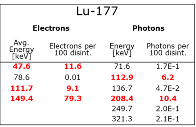
Table 1: Frequency and energy of the particles emitted at the decay of Lu-177 or its daugther nuclides
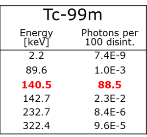
Table 2: Frequency and energy of the particles emitted at the decay of Tc-99m or its daugther nuclides
When imaging these radionuclides with SPECT, one measures only the gamma particles that are emitted either at the decay site, or those ejected by the deceleration of emitted beta particles. The gamma spectra for Tc-99m and Lu-177 are therefore substantially different. Figure 1 shows the gamma spectrum of Lu-177 measured with a high purity germanium detector.
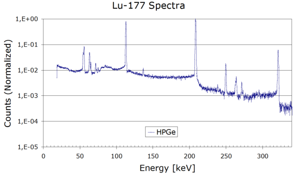
Figure 1: Energy spectrum of the photons emitted by Lu-177, measured with a high-purity Germanium detector.
As a consequence, the correction for scattered radiation is more difficult for Lu-177, compared to Tc-99m. At the very least, one needs to apply an upper- and lower-scatter window, as seen as in Figure 2.
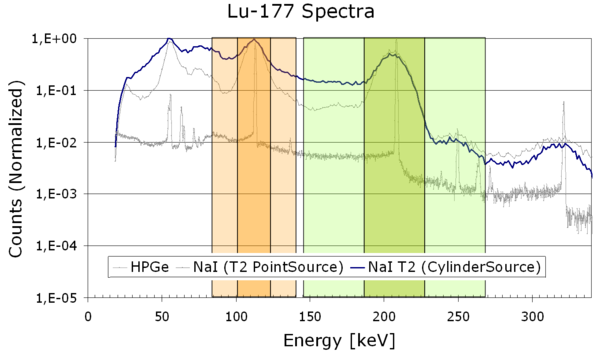
Figure 2: Photopeak windows (dark green & dark orange) and upper/lower scatter windows (light colors) for Lu-177
Please see the section “Scatter” of the project “Absolute Quantification with SPECT/CT” for information why scatter correction is necessary and how it works.
Problem statement: effective imaging resolution
Due to the higher energy photons encountered in Lu-177 SPECT, one needs to use medium energy (ME) collimators, as opposed to the low energy (LE) collimators for Tc-99m. Among other effects, these collimators typically yield a reduced spatial resolution. For illustrating this, we imaged point sources of Tc-99m and Lu-177 using LE and ME collimation, respectively. Both sources were taken at the same collimator-to-source distance. According to Figure 3, we find a broader point spread function (PSF) for Lu-177 and measured a corresponding wider FWHM.
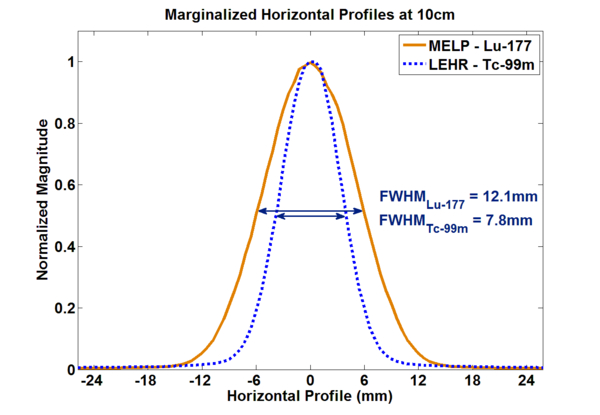
Figure 3: Line profiles trough Lu-177/Tc-99m point sources. Courtesy of  J. Sanders
Problem statement: optimal reconstruction parameters
Mainly as a consequence of the differing imaging resolution, the convergence properties of the iterative OSEM reconstruction of the Lu-177 SPECT images are slightly changed compared to those of the Tc-99m reconstructionThe relevant parameters of reconstructed SPECT images, besides geometrical correctness, are the amount of recovery (e.g. expressed as the ratio of measured to true activity concentration), the amount of noise (e.g. expressed as the standard deviation normalized by the measured activity concentration of a volume of interest), and the absence of other unwanted artefacts (e.g. image ringing). For the evaluation of suitable reconstruction parameters, we performed phantom acquisitions using spheres that were filled with a known amount of radioactivity and which were embedded in a filled cylinder that also contained a certain amount of radioactivity as a background. The experiment should mimic a typical patient acquisition where one often encounters lesions with a high amount of radioactive uptake in a background that has a lower amount of (often unspecific) uptake. Figure 4 shows the recovery of the activity concentration (as a fraction of the true concentration) in relation to the reconstruction parameters. Only results from the largest spheres (16 ml, 8 ml) are shown.
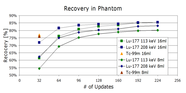
Figure 4: Recovery coefficients for Tc-99m and Lu-177 in a phantom with hot spheres, in dependency of the number of updates in the iterative reconstruction, separately for the two photopeaks
We find that the recovery rises with increasing number of updates of the OSEM reconstruction and saturates at some point. The recovery for Tc-99m is higher than for Lu-177, which is due to the higher spatial resolution of Tc-99m SPECT. The recovery saturates at around 85%, which is a limit imposed by partial volume effects. It can be seen here that without any further corrections, the maximally achievable recovery depends on the size of the spheres. The recovery is lower for smaller structures.For standard OSEM, without any further regularization, an increasing number of updates also increases the image noise, which is shown in Figure 5. The optimal number of updates for quantitative Lu-177 reconstruction is consequently a compromise between recovery and noise. Since we focus on correct quantitation for a potential application in dosimetry and not on clinical reading by physicians, we chose the optimal point to achieve correct recovery values, accepting the increased noise levels.
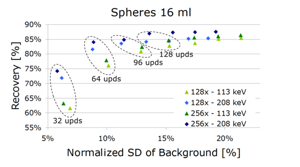
Figure 5: Bias (recovery) vs. image noise (SD) for reconstructions with a varying number of updates of the 16 ml sphere, separately for the two photopeaks
Validation: phantom experiments
For the calibration of our camera system, we used the calibration procedures outlined in the section “Calibration” of the project “Absolute Quantification with SPECT/CT”. We found a calibration factor of 0.61 cpm/kBq for the 208 keV photopeak and 0.65 cpm/kBq for the 113 keV photopeak for our SPECT/CT system (Siemens Symbia T2, 3/8” crystal, MELP collimators).
Validation: patient measurements
Using the aforementioned calibration factors, we determined the radioactivity concentration in the urinary bladder in attenuation and scatter corrected SPECT/CT images and compared it to the concentration in urine samples measured in a well counter. We found an average error of 8.0%. The size of the VOIs was on average 111 ml. This shows that SPECT/CT can be considered as quantitatively accurate in similarly sized VOIs. These quantitative results, as well as the 3-D nature of SPECT imaging, encourage the use of SPECT/CT for the dosimetry of Lu-177 compounds.
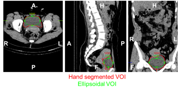
Figure 6: Hand segmented and ellipsoidal volumes of interest in the urinary bladder of a patient for the validation study