
Contact

 +49-9131-85-27775
 +49-9131-85-27270
Secretary
| Monday | 8:00 - 12:15 |
| Tuesday | 8:00 - 16:45 |
| Wednesday | 8:00 - 16:45 |
| Thursday | 8:00 - 16:45 |
| Friday | 8:00 - 12:15 |
Address
Lehrstuhl für Informatik 5 (Mustererkennung)
Martensstr. 3
91058 Erlangen
Germany
Powered by

Image Analysis
The Image Analysis Group is dedicated to extract information from images. Examples are the outlining of specific structures in 2D and 3D images, like extraction of pages in CT scans of books or the detection of lesions in mammographic images.
Place
Usually the colloquium takes place in room RZ 2.009 (conference room). If the room is not available, you can find us either in e-Studio (RZ 2.037) or in our own seminar room 01.134.
Next sessions
| Date | Responsible Person | Speaker | Type & Title of Contribution |
|---|---|---|---|
04.06.2019 |
Viktor Haase |
Improving FISTA: Faster, Smarter and Greedier | |
28.05.2019 29.01.2019 | Weilin Fu Lennart Husvogt | MONet: Unsupervised Scene Decomposition and Representation OCTA Image Generation | |
| 05.02.2019 | Viktor Haase | Exploring the Space Between Smoothed and Non-Smooth Total Variation for 3-D Iterative CT Reconstruction | |
| 26.02.2019 | Weilin Fu |  Fast End-to-End Trainable Guided Filter Fast End-to-End Trainable Guided Filter | |
| 12.03.2019 | Mario Amrehn | Robot User Comparison and Understanding Disentangling in β-VAE | |
| from 12.2. | to 23.4.2019 | no lectures, colloquium on demand (e.g. thesis talks) | |
Mailing list subscription management page for students and guests.
In case a remote participation is needed, please  contact the organizer of the colloquium.
Running Projects
 |
3-D Reconstruction of Historical DocumentsThis project focuses on the reconstruction of historical documents that can not be opened or page-turned anymore. We perform X-ray CT scans and develop algorithms to extract and visualize pages from the 3-D volume. This is possible because historical documents were commonly written with ancient inks consisting of metallic particles. |
 |
Parameter-Optimization for DBT Imaging SystemsDBT can be used for screening of the human breast which implies a specific demand of best possible image quality w.r.t. diagnostic value. One diagnostic measure is the detectability of lesions which is connected with the concept of model observers. Ultimately, we use these kinds of measure to optimize DBT reconstruction parameters. |
 |
Image Quality Assessment with Human ObserversThe penalized least-square reconstruction with statistical weights is a popular model-based iterative reconstruction method for CT. In our project we examine the impact of the statistical weights on image quality. We conduct human observer studies to test the lesion detection performance on simulated phantom data. The final image quality assessment is based on the analysis of the localization receiver operating characteristic. |
 |
Airway SegmentationAirway Segmentation is a challenging task, because the radius range of the airway can be large, and image quality can be bad. We are using differen methods to segment airway out, including region growing, Frangi tubeness, cavity enhancement filtering, circle detection filtering, gradient vector flow, and super pixel. |
 |
Image Procesing for Ophthalmic Optical Coherence TomographyOptical Coherence Tomography is a widely used imaging modality in ophthalmology. Ocular diseases such as diabetic retinopathy and age-related macular degeneration are becoming more common due to the increasing prevalence of diabetes and increasing life expectancies in the population. This project aims to aid in the diagnosis and mointoring of ophthalmic dieases. |
Finished Projects
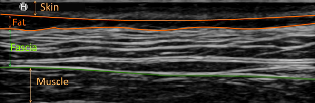 |
Segmentation of Fat and Fascia Layers in Ultrasound ImagesOne recent research area in zoology is to measure the thickness of the fat and fascia layer within ultrasound images. Our goal is to develop an algorithm for fully automatic separation of those layers. Furthermore, we want to provide a GUI for specialist such that there is no more need to manually measure the layers. |
 |
Machine Learning-based Material Decomposition for Spectral X-ray ImagingBenefiting from multi-energy X-ray imaging technology, material decomposition facilitates the differentiation of different materials in X-ray imaging. We propose a novel machine learning-based pipeline to perform material decomposition using machine learning algorithms. Feature extraction is involved into the pipeline to improve the performance of material decomposition. |