Contact

 +49-9131-85-27775
 +49-9131-85-27270
Secretary
| Monday | 8:00 - 12:15 |
| Tuesday | 8:00 - 16:45 |
| Wednesday | 8:00 - 16:45 |
| Thursday | 8:00 - 16:45 |
| Friday | 8:00 - 12:15 |
Address
Lehrstuhl für Informatik 5 (Mustererkennung)
Martensstr. 3
91058 Erlangen
Germany
Powered by

Medical Image Segmentation
Medical Image Segmentation is the process of automatic or semi-automatic detection of boundaries within a 2D or 3D image. A major difficulty of medical image segmentation is the high variability in medical images. First and foremost, the human anatomy itself shows major modes of variation. Furthermore many different modalities (X-ray, CT, MRI, microscopy, PET, SPECT, Endoscopy, OCT, and many more) are used to create medical images. The result of the segmentation can then be used to obtain further diagnostic insights. Possible applications are automatic measurement of organs, cell counting, or simulations based on the extracted boundary information.
Research Directions

Vessel Segmentation and Blood Flow
Segmentation of cerebral vessels and subsequent use for numerical blood flow simulations.
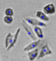
Cell Detection
Detection of cells in bright-field microscope images.

Coronary Vessel Segmentation
Segmentation and modelling of the coronary vessel tree in intra-procedural data for quantitative measurements.

Personalized Segmentation of the Heart Great Vessels and Valves
Developing patient-specific cardiac models, estimated from available multi-modal images, to enable advanced clinical applications for the management of cardiovascular disease.

Analysis of Resting State Functional Magnetic Resonance Images
Detection of patient characteristics from resting state functional magnetic resonance images, with a focus the early diagnosis of Alzheimer's disease.