
Contact


Address
Universität Erlangen-Nürnberg
Chair of Computer Science 5 (Pattern Recognition)
Martensstrasse 3
91058 Erlangen
Germany
Chair of Computer Science 5 (Pattern Recognition)
Martensstrasse 3
91058 Erlangen
Germany
Powered by

Dr.-Ing. Christopher Rohkohl
Alumnus of the Pattern Recognition Lab of the Friedrich-Alexander-Universität Erlangen-Nürnberg
The vision of my work is to develop methods that open up the possibility to visualize highly dynamic (moving) vital parts of the human body as the heart. This provides one key towards the development of novel diagnostic tools and medical treatment procedures.
Introduction to my basic research: Interventional Imaging using C-arm Systems
During the recent years 3-D imaging has become a standard tool for diagnosis and planning
in the clinics using well-known imaging modalities as CT or MRT. The integration of those
image during an intervention is difficult and requires various registration techniques.
A solution that has emerged in recent years is the use of fluoroscopic C-arm systems which
are highly common in the interventional field for reconstructing 3-D images.
The workflow can be seen in the following three videos from one of our clinical cooperations (Klinikum Coburg GmbH):
The general goal of my research topics is to enrich that workflow for mainly cardiac applications by providing high quality, fast and reliable algorithms for
the 3-D reconstruction of challenging subjects.
Of course this work is highly interdisciplinary and requires techniques and concepts from the complete field of medical image processing.
In the following an overview of the detailed research topics is provided.

Research Project: Non-Periodic Motion Estimation and Motion Compensation Algorithms
The primary goal of our research activities in this project is the development of methods for estimating and correcting
cardiac motion in order to increase the image quality of cardiac vasculature reconstruction.
In interventional environments patients often do have arrhythmic heart signals or cannot hold
breath during the complete data acquisition. This important group of patients
cannot be reconstructed with current approaches that do strongly
depend on a high degree of cardiac motion periodicity for working properly.
In this project we try to develop novel algorithmic approaches to cardiac vasculature reconstruction and therefore address
the following questions:
Read More ...
Keywords: Motion compensation, dynamic 3-D reconstruction, cardiac imaging, C-arm CT
- Development of algorithms for motion estimation and reconstruction without periodicity assumption or ECG information.
- Analysis and development of models for the description of cardiac and breathing motion.
- Development of algorithms for estimatating the optimal heart phase for phase-correlated reconstruction algorithms.
- Development of methods for the segmentation and functional assessment from 3-D plus time image data.
Read More ...
Keywords: Motion compensation, dynamic 3-D reconstruction, cardiac imaging, C-arm CT
Research Project: CAVAREV - CArdiac VAsculature Reconstruction EValuation
The main goal of the CAVAREV-project is to enable an easy and objective comparison of different dynamic reconstruction algorithms.
The area of application is the 3-D and 4-D reconstruction of contrasted cardiac vessels, e.g. the coronary arteries using C-arm CT (rotational angiography).
Various methods exist in literature with lots of nice results. However, the results can vary significantly between phantom and real clinical data.
Therefore, CAVAREV was developed, which provides:
Keywords: Motion compensation, motion estimation, Benchmark, Coronary reconstruction
- Open (i.e. public) and easy accessible projection data.
- Anatomically and physiologically correct projection data based on patient data.
- Two degrees of difficulty: (a) Strictly periodic cardiac motion and (b) an aperiodic combination of cardiac and breathing motion.
- An online evaluation platform where 3-D reconstructions can be uploaded and the reconstruction quality is assessed objectively.
For more information please visit  www.cavarev.com.
Keywords: Motion compensation, motion estimation, Benchmark, Coronary reconstruction
Research Project: RabbitCT - An Open Platform for Benchmarking 3-D Cone-Beam Reconstruction Algorithms
In this project we consider the problem of evaluating hardware acceleration schemes for 3-D cone beam reconstruction algorithms.
Researchers and industry work hard on hardware-optimized 3D reconstruction which is crucial for a seamless workflow integration.
A crucial limitation, however, of these publications is that the presented results are not comparable to each other.
This is mainly due to variations in data acquisitions, preprocessing, and chosen geometries and the lack of a common publicly available test dataset.
With RabbitCT we provide such a standardized dataset that allows for substantial comparison of hardware accelerated backprojection methods.
In summary, it is an online platform for worldwide comparison in reconstruction performance and ranking on different architectures using a
specific high resolution C-arm CT dataset of a rabbit. This includes a sophisticated benchmark interface, a prototype implementation in C++, and image quality measures.
Keywords: Benchmark testing, C++ language, computer graphics, computerised tomography, image reconstruction, medical image processing
For more information please visit www.rabbitct.com.
Keywords: Benchmark testing, C++ language, computer graphics, computerised tomography, image reconstruction, medical image processing
Research Project: Image-Based Gating
Applying the concept of retrospective electrocardiogram gating (ECG) to the acquisition of
multiple, ECG-triggered rotational acquisitions using a C-arm system allows the 3D+t reconstruction of the
heart. The process of retrospective gating is a crucial component of 3-D reconstruction. The gold-standard
in gating is still ECG based. However, the ECG signal does not directly reflect the mechanical situation of
the heart. Therefore an alternative gating method, based on the acquired projection data is required. Our
goal is to provide an image-based gating (IBG) method without ECG such that already acquired projection
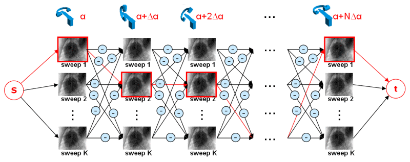
data from a multi-sweep acquisition can still be used for reconstruction. We formulate the gating problem as a
shortest-path optimization problem. All acquired projection images build a directed graph and the path costs are
defined by projection image similarities that are based on image metrics to measure the heart phase similarity.
The optimization is additionally regularized to prefer solutions where the path segment of consecutive selected
projections acquired along a particular forward or backward C-arm sweep is short. This regularization depends
on an estimated average heart rate that is also estimated using an image-based method. First promising results
using in-vivo data are presented and compared to standard ECG gating.
Read More ...
Keywords: Cardiac C-arm CT, Retrospective Gating, Electrocardiogram-Based Gating, Image-Based Gating
Keywords: Cardiac C-arm CT, Retrospective Gating, Electrocardiogram-Based Gating, Image-Based Gating