Contact


Address
Chair of Computer Science 5 (Pattern Recognition)
Martensstrasse 3
91058 Erlangen
Germany
Powered by

Dr.-Ing. Alexander Brost
Alumnus of the Pattern Recognition Lab of the Friedrich-Alexander-Universität Erlangen-Nürnberg
Cryo-Balloon Reconstruction from Two Views
Andreas Kleinoeder,  Alexander Brost, Felix Bourier,
  Martin Koch, Klaus Kurzidim, Joachim Hornegger, and Norbert Strobel
Atrial fibrillation (AFib), the most common sustained arrhythmia, is a major cause of stroke. One treatment option for AFib is the electrical isolation of the four pulmonary veins attached to the left atrium by catheter ablation. It is recommended, e.g., when patients fail drug therapy. C-arm X-ray systems are used for AFib procedures. Augmented fluoroscopy using a perspectively forward projected overlay representation of 3-D objects onto live fluoroscopic images has become a useful tool procedure for navigation when performing ablation procedures. Using a modern biplane C-arm system with two imaging planes, not only visualization but also 3-D localization and object reconstruction has become possible. Two risk factors of the current radio-frequency catheter ablation approach are pulmonary vein stenosis and esophageal fistula. To reduce these risks, cryo-balloon catheter ablation techniques can be used. Unfortunately, current navigation tools do not provide tools to localize and visualize cryo-balloon catheters in 3-D. In our approach, the catheter is modeled as a sphere in 3-D. Methods to reconstruct ellipsoids either require three views or additional 3-D information. A method for sphere reconstruction has already been proposed, but it turns out to be rather sensitive to noise. This is why we present a novel method to reconstruct a cryo-balloon catheter from two views. It is well suited to compute a 3-D model even in the presence of noise. Used as part of a fluoroscopic overlay image for augmented fluoroscopy applications, we expect that our method can further increase the safety and effectiveness of the cryo-balloon ablation approach.
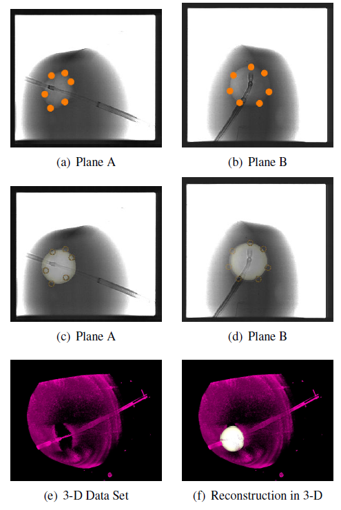
The reconstruction process consists of two steps. In the first step, the 2-D input data is processed and in the second step, the 3-D catheter model is generated. As input, manually selected 2-D points on the boundary of the balloon-catheter in both views are needed.
Details can be found here.
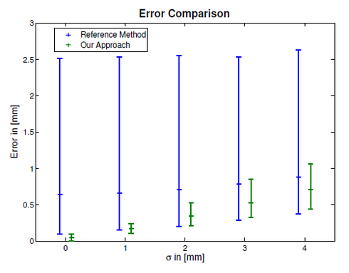 |
For evaluation of our method, we performed a simulation study and performed experiments using a real biplane C-arm X-ray system. For the simulation, we computed 250 spheres each at a different positions in 3-D space within a maximum distance to the center of the volume of 150 mm and with a radius between 5 mm and 15 mm. The simulation was performed using ideal C-arm projection matrices as described here and here. The C-arm positions were chosen to be 90 degrees apart which is similar to a clinical setup. We compared our approach to the reference method in the presence of Gaussian noise with a standard deviation of 0 mm to 4 mm. The results are shown in Figure 1. The experiments were performed on an Artis zee biplane C-arm X-ray system (Siemens AG, Healthcare Sector, Forchheim, Germany) at a clinical partner site (Krankenhaus Barmherzige Brueder, Regensburg, Germany). We used a cryo-ballon catheter placed in a bucket filled with contrast agent to evaluate our 3-D sphere reconstruction from two views. At first, a C-arm computed tomography data set (syngo DynaCT, Siemens AG, Forchheim, Germany) was acquired to obtain a 3-D data set of the object. Afterwards, biplane fluoroscopic images were taken, the 2-D input points for our algorithm were manually selected, and sphere reconstruction from two views was performed.
The reconstruction result was visually compared to the 3-D data set, see Fig. 2. The associated 2-D images are shown in Fig. 1. During the course of this work, we found our sphere reconstruction method from two views robust and easy to use. We contribute the better performance of our method to two facts. First, we do not use principal component analysis (PCA) for ellipse fitting. PCA is of advantage if many samples are present that facilitate a robust parameter estimate. In our case, we only require a few points set along object boundaries. Usually five to seven points are sufficient. In such a case, the PCA approach turns out to be very sensitive especially if the samples are not equally distributed along the object boundary. Second, we do not rely on only one or two frontier points, as proposed for the reference method. Instead, we use about 100 points to determine the radius of the sphere in 3-D. Thanks to these improvements, our 3-D reconstruction is more robust to noise. The proposed method is likely to be helpful during cryoballoon catheter ablation procedures as it provides visual feedback to the physician, e.g., about previous balloon positions when multiple freezing treatments are applied to the same pulmonary vein. In the next steps, we are going to clinically evaluate the proposed approach to get a better understanding of its utility for cryo-balloon catheter treatments. We expect that the use of our approach will further improve the safety and efficacy of this treatment option.
 |
Full paper available here.