
Contact


Address
Chair of Computer Science 5 (Pattern Recognition)
Germany
Powered by

Dipl.-Inf. Andreas Wimmer
Alumnus of the Pattern Recognition Lab of the Friedrich-Alexander-Universität Erlangen-Nürnberg
Organ Segmentation for Perfusion CT
My research project consists in the development and evaluation of methods for abdominal organ segmentation in perfusion Computed Tomography (Perfusion CT) images.
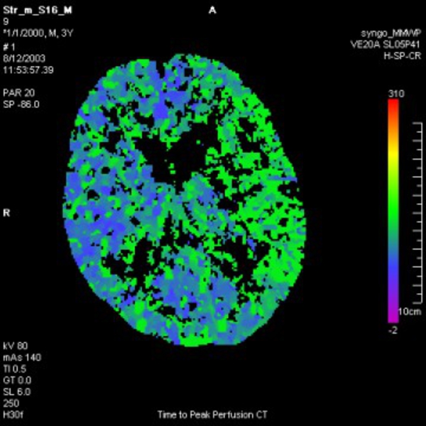
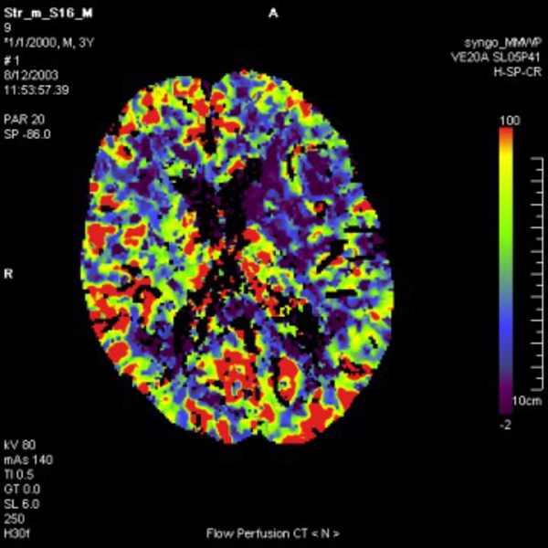
Over the last years, perfusion imaging has become an important diagnostical tool for patients suffering from strokes or tumors. In the context of stroke, perfusion is reduced for affected areas, whereas in the context of tumors, perfusion is increased, a result of the tumor neovascularization.
In perfusion CT, a contrast agent is injected into the blood supply for the region of interest. Slice images are acquired with a temporal resolution of about 1 to 3 seconds and features like time-to-peak of contrast enhancement, blood flow, or blood volume are calculated.
For brain perfusion CT, algorithms exist which remove surrounding tissues and bone structures, thereby allowing the analysis to be restricted to the region of interest.
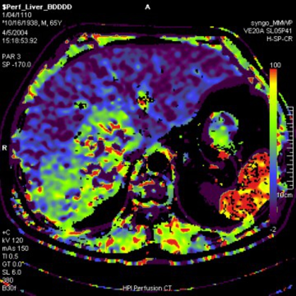
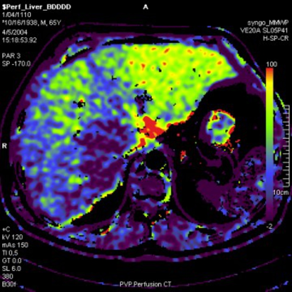
The situation is a lot more complicated for abdominal perfusion CT. Organs like the liver or the pancreas exhibit a high variability in shape and are surrounded by tissues with similar intensities, requiring sophisticated segmentation algorithms. Additionally, organ movements and deformations, for example due to respiratory motions, have to be compensated through image registration in order to obtain an accurate perfusion analysis.
Figure 1: Brain perfusion CT
 |
 |
 |
 |
The situation is a lot more complicated for abdominal perfusion CT. Organs like the liver or the pancreas exhibit a high variability in shape and are surrounded by tissues with similar intensities, requiring sophisticated segmentation algorithms. Additionally, organ movements and deformations, for example due to respiratory motions, have to be compensated through image registration in order to obtain an accurate perfusion analysis.
Figure 2: Liver perfusion CT
 |
 |
 |
 |
The challenge is to develop image processing methods which are fast, accurate, and robust, and require only a minimum amount of user interaction, in order to meet the requirements for clinical work flows.
This research project is supported by our industrial partner  Siemens Medical Solutions.