Contact


Address
Chair of Computer Science 5 (Pattern Recognition)
Germany
Powered by


Dipl.-Inf. Andreas Wimmer
Alumnus of the Pattern Recognition Lab of the Friedrich-Alexander-Universität Erlangen-Nürnberg
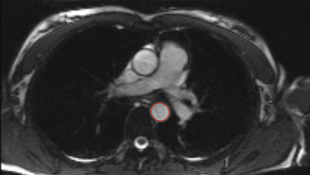
Aorta Segmentation for Interventional MRI
The vision of this project is to provide visual guidance through a virtual endoscope. Interventions like stent placement are frequently carried out using guidance form plain X-ray or MRI images. However, knowing the exact anatomy and being able to freely position the point of view can be extremely valuable during an intervention. We therefore developed a parametric model of the human aorta. In combination with our fast edge-based level set active contour operating on cross-sectional images, segmentation and visualization is possible at interactive rates for a large number of slices. The principle is illustrated through Fig 1. |
|
Fig. 1. Virtual endoscopy. Segmentation of the aortic cross-section for several slices. A 3-D model is generated in real-time from the boundary and allows the physician to freely position the point of view inside or outside the vessel. The deformation of the aorta due to the blood flow is clearly visible.
|