
Contact


Address
Chair of Computer Science 5 (Pattern Recognition)
Germany
Powered by

Dr.-Ing. Michael Manhart
Alumnus of the Pattern Recognition Lab of the Friedrich-Alexander-Universität Erlangen-Nürnberg
Interventional 4-D Tissue Perfusion Imaging with C-arm CT
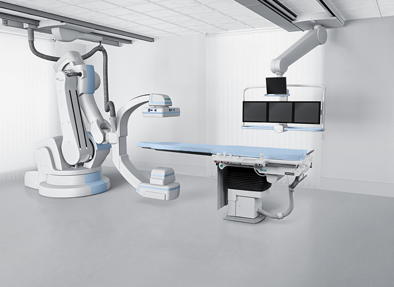
In this project, we investigate the use of a C-arm angiography system capable of CT-like imaging (C-arm CT, Fig. 1) to measure tissue perfusion. Tissue perfusion information, such as cerebral blood flow (CBF), can be used for stroke diagnosis and to guide further stroke treatment. While CT and MR-based perfusion imaging is well established perfusion imaging in the interventional suite using a C-arm CT is not available yet.
Current catheter-guided stroke therapy procedures such as intra-arterial thrombolysis allow to potentially reperfuse salvageable ischemic tissue. For this purpose the patient is transported to an interventional suite with an C-arm angiography system. Perfusion measurement using C-arm systems would allow assessing the perfusion parameters directly before and during the interventional procedure and help to determine the treatment success and endpoint.
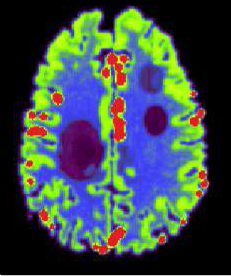
During perfusion imaging a region of interest is imaged several times at short intervals to study the flow of a previously injected bolus of contrast agent (Fig. 2). Using a C-arm CT for perfusion imaging faces certain challenges. In contrast to conventional CT scanners, the acquisition time for one data set is much longer (3-5 seconds compared to less than 1 second in CT) and thus the sample rate for the contrast flow is comparably low. Furthermore, inconsistencies in the projection images - due to the changing attenuation values from the contrast agent during the acquisition time - may cause reconstruction artifacts. Also the peaks of the contrast flow curves typically lie in a range of 5-30 HU, thus perfusion imaging is very sensitive to noise.
This project is in collaboration with:
- Siemens AG, Healthcare Sector, Angiography & Interventional X-Ray Systems
- Department of Neuroradiology, Universitätsklinikum Erlangen (Prof. Dr. med. Arnd Dörfler)
- Department of Radiology, Stanford University, USA (Prof. Rebecca Fahrig)
 |
 |
 |
The focus on our current research is in dynamic interative reconstruction algorithms for perfusion C-arm CT to deal with the challenges of slow C-arm rotation speed and low contrast-to-noise level in the brain tisse contrast flow.
The novel algorithm reconstructs TACs described by a weighted sum of linear spline functions. To reduce noise a novel regularization technique based on joint bilateral filtering is applied.
For evalutation of the algorithms a digital brain perfusion phantom was created, which can be downloaded here.
Cone-Beam Reconstruction with Displaced Flat Panel Detector
M Manhart, F Dennerlein and H Kunze: "ONLINE CONE BEAM RECONSTURCTION WITH DISPLACED FLAT PANEL DETECTOR", First CT Meeting, pp. 53-56, Salt Lake City, UT, 2010.
Respiratory Motion Compensation for Fluoroscopic Coronary Roadmapping
M Manhart, Y Zhu and D Vitanovski: "SELF-ASSESSING IMAGE BASED MOTION COMPENSATION FOR FLUOROSCOPIC CORONARY ROADMAPPING", pp. 1065-1069, IEEE ISBI, Chicago, IL, 2011. (Top10 finalist in the ISBI 2011 student paper competition)