Friedrich-Alexander-Universität Erlangen
Lehrstuhl für Mustererkennung
Martensstraße 3
91058 Erlangen

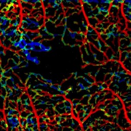
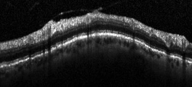
In the recent decade, ophthalmic imaging has proven to be a steadily growing field of research. A milestone in ophthalmology was the transfer of Optical Coherence Tomography (OCT) from research to clinical use. Compared to conventional fundus photography, OCT allows the three dimensional, depth resolved visualization of the human retina, while preserving the non-invasiveness. Recently, a radical change occured in the field of OCT research with the clinical introduction of OCT angiography (OCTA), which further adds dye-free imaging of the underlying vasculature.
To facilitate an efficient analysis of this vastly increased amount of information, new processing algorithms are required to support the treating clinician. Our research focus is twofold: On the one hand, we develop advanced motion compensation and signal reconstruction algorithms to achieve artifact-free OCT(A) signals of best possible quality. On the other hand, we develop informative visualization tools and novel image analysis and segmentation methods. By combining both efforts, we want to improve the understanding of the most prevalent eye diseases, allowing for more accurate treatment and thus improved patient outcome.
To enable research with state-of-the-art technology while preserving a close link to the clinical needs, the work of our group is embedded in a multidisciplinary environment including optical engineers at the ![]() Massachusetts Institute of Technology, Cambridge, USA and clinicians at the
Massachusetts Institute of Technology, Cambridge, USA and clinicians at the ![]() New England Eye Center, Boston, USA and the Department of Ophthalmology at the
New England Eye Center, Boston, USA and the Department of Ophthalmology at the ![]() University Clinic Erlangen.
University Clinic Erlangen.

The target of this project is to explore the benefits of the OCTA signal in a wide range of applications. These range from visualization and analysis of the OCTA signal alone, up to extending previously OCT-only algorithms to hybrid OCT-OCTA variants that exploit the additional information of the OCTA signal. Examples are:
Motion Correction
Signal Enhancement
Layer Segmentation
Artifact Correction
Disease Classification

 Advanced fundus imaging for early detection of eye diseases
 Layer segmentation e.g. retinal nerve fiber segmentation (ARVO 2008 Poster
 ![]() PDF)
PDF)
 Denoising (Poster ARVO 2010 Poster
 ![]() PDF)
PDF)
 Automated glaucoma detection (ARVO 2009 Poster
 ![]() PDF)
PDF)

