Dr.-Ing. Johannes Feulner
Alumnus of the Pattern Recognition Lab of the Friedrich-Alexander-Universität Erlangen-Nürnberg
Estimating the visible body portion of CT volume images
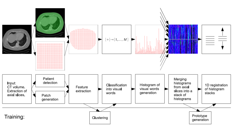
Being able to automatically determine which portion of the human body is shown by a CT volume image offers various possibilities like automatic labeling of images or initializing subsequent image analysis algorithms. We developed a method that takes a CT volume as input and outputs the vertical body coordinates of its top and bottom slice in a normalized coordinate system whose origin and unit length are determined by anatomical landmarks. Each slice of a volume is described by a histogram of visual words: Feature vectors consisting of an intensity histogram and a SURF descriptor are first computed on a regular grid and then classified into the closest visual words to form a histogram. The vocabulary of visual words is a quantization of the feature space by offline clustering a large number of feature vectors from prototype volumes into visual words (or cluster centers) via the K-Means algorithm. For a set of prototype volumes whose body coordinates are known the slice descriptions are computed in advance. The body coordinates of a test volume are computed by a 1D rigid registration of the test volume with the prototype volumes in axial direction. The similarity of two slices is measured by comparing their histograms of visual words.


