
Contact
 +49 9131 85 28982
 +49 9131 85 27270

Address
Chair of Computer Science 5 (Pattern Recognition)
Martensstr. 3
91058 Erlangen
Germany
Powered by

Dr.-Ing. Oliver Taubmann M. Sc.
Alumnus of the Pattern Recognition Lab of the Friedrich-Alexander-Universität Erlangen-Nürnberg
Projects
Oliver Taubmann, Günter Lauritsch, Gregor Krings, Andreas Maier
-
Dynamic cardiac imaging typically requires the use of gating. In the case of computed tomography (CT), this results in an angular undersampling that renders reconstruction difficult. Prior work has shown that incorporating information from the full scan, i.e. from all cardiac phases, can be beneficial in this regard, for instance by regularization. This paper compares three convex temporal regularizers for 4-D cardiac C-arm CT in both a numerical phantom and clinical patient data. Regularizations based on the nuclear norm, temporal total variation as well as a tight-frame wavelet transform are studied. While all of them improve reconstruction quality notably, the former turns out to be the least effective. The latter two yield comparable results at near-optimal parameterization. However, temporal total variation appears to be more forgiving w.r.t. over-regularization.
Articles in Conference ProceedingsProceedings of the 4th International Conference on Image Formation in X-ray Computed Tomography (4th International Conference on Image Formation in X-ray Computed Tomography), Bamberg, Germany, 18.07.-22.07.2016, pp. 545-548, 2016 (BiBTeX, Who cited this?)
Oliver Taubmann, Günter Lauritsch, Andreas Maier, Rebecca Fahrig, Joachim Hornegger
-
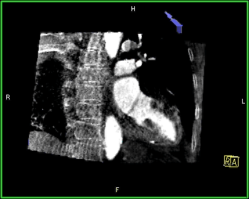
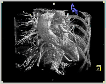
Detailed analysis of cardiac motion would be helpful for supporting clinical workflow in the interventional suite. With an angiographic C-arm system, multiple heart phases can be reconstructed using electrocardiogram gating. However, the resulting angular undersampling is highly detrimental to the quality of the reconstructed images, especially in non-ideal intraprocedural imaging conditions. Motion-compensated reconstruction has previously been shown to alleviate this problem, but it heavily relies on a preliminary reconstruction suitable for motion estimation. In this work, we propose a processing pipeline tailored to augment these initial images for the purpose of motion estimation, and assess how it affects the final images after motion compensation.
The following combination of simple, direct methods inspired by the core ideas of existing approaches proved beneficial: (a) Streak reduction by masking high-intensity components in projection domain after filtering. (b) Streak reduction by subtraction of estimated artifact volumes in reconstruction domain. (c) Denoising in spatial domain using a joint bilateral filter guided by an uncompensated reconstruction. (d) Denoising in temporal domain using an adaptive Gaussian smoothing based on a novel motion detection scheme.
Experiments on a numerical heart phantom yield a reduction of the relative root-mean-quare error from 89.9% to 3.6% and an increase of correlation with the ground truth from 95.763% to 99.995% for the motion-compensated reconstruction when our processing is applied to the initial images. In three clinical patient data sets, the signal-to-noise ratio measured in an ideally homogeneous region is increased by 37.7% on average. Overall visual appearance is improved notably and some anatomical features are more readily discernible.
Our findings suggest that the proposed sequence of steps provides a clear advantage over an arbitrary sequence of individual image enhancement methods and is fit to overcome the issue of lacking image quality in motion-compensated C-arm imaging of the heart. As for future work, the obtained results pave the way for investigating how accurately cardiac functional motion parameters can be determined with this modality.
Supplementary animated images can be found here.
Journal ArticlesMedical Physics, vol. 43, no. 2, pp. 883-893, 2016 (BiBTeX, Who cited this?)
Oliver Taubmann, Günter Lauritsch, Andreas Maier, Rebecca Fahrig, Joachim Hornegger
-
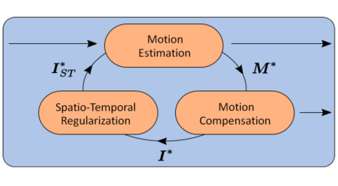
C-arm computed tomography reconstruction of multiple cardiac phases could provide a highly useful tool to interventional cardiologists in the catheter laboratory. Today, however, for clinically reasonable acquisition protocols the achievable image quality is still severely limited due to undersampling artifacts. We propose an iterative optimization scheme combining image registration, motion compensation and spatio-temporal regularization to improve upon the state-of-the-art w.r.t. image quality and accuracy of motion estimation. Evaluation of clinical cases indicates an improved visual appearance and temporal consistency, evidenced by a strong decrease in temporal variance in uncontrasted regions accompanied by an increased sharpness of the contrasted left ventricular blood pool boundary. In a phantom study, the universal image quality index proposed by Wang et al. is raised from 0.80 to 0.95, with 1.0 corresponding to a perfect match with the ground truth. The results lay a promising foundation for interventional cardiac functional analysis.
Supplementary animated images can be found
 here.Articles in Conference ProceedingsMedical Image Computing and Computer-Assisted Intervention – MICCAI 2015, Munich, Germany, 05.10-09.10.2015, pp. 579-586, 2015, ISBN 978-3-319-24571-3 (BiBTeX, Who cited this?)
Oliver Taubmann, Günter Lauritsch, Andreas Maier, Rebecca Fahrig, Joachim Hornegger
-
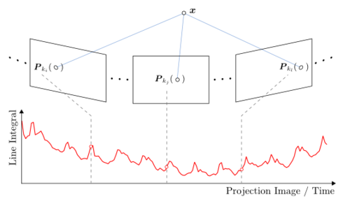
Time-resolved 3-D cardiac imaging in the catheter laboratory using C-arm computed tomography (CT) could provide valuable information to physicians during interventions. However, for clinically reasonable acquisition protocols, electrocardiography (ECG) gated reconstructions of individual heart phases are severely degraded due to sparse view sampling artifacts. As they appear at different locations for each heart phase, these artifacts strongly influence motion estimation, thus impairing a subsequent motion-compensated reconstruction which has been shown to greatly improve image quality. We present a method for reducing such artifacts by an adaptive smoothing in the temporal domain, guided by a projection-based motion detection step which incorporates the heart rate as prior knowledge. In our experiments on clinical data and a numerical heart phantom, we show that the increased temporal consistency achieved for initial images propagates into the motion-compensated reconstructions, improving their quality.
Articles in Conference ProceedingsProceedings of the 13th International Meeting on Fully Three-Dimensional Image Reconstruction in Radiology and Nuclear Medicine (13th International Meeting on Fully Three-Dimensional Image Reconstruction in Radiology and Nuclear Medicine), Newport, Rhode Island, USA, 31.05.2015-04.06.2015, pp. 532-535, 2015 (BiBTeX, Who cited this?)
Oliver Taubmann, Jens Wetzl, Günter Lauritsch, Andreas Maier, Joachim Hornegger
-
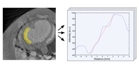
Organ motion occuring during acquisition of medical images can cause motion blur artifacts, thus posing a major problem for many commonly employed modalities. Therefore, compensating for that motion during image reconstruction has been a focus of research for several years. However, objectively comparing the quality of different motion compensated reconstructions is no easy task. Often, intensity profiles across image edges are utilized to compare their sharpness. Manually positioning such a profile line is highly subjective and prone to bias. Expanding on this notion, we propose a robust, semi-automatic scheme for comparing edge sharpness using an ensemble of profiles. We study the behavior of our approach, which was implemented as an
open-source tool, for synthetic data in the presence of noise and artifacts and demonstrate its practical use in respiratory motion-compensated MRI as well as cardiac motion-compensated C-arm CT.Articles in Conference ProceedingsBildverarbeitung für die Medizin 2015, Lübeck, 15.03-17.03, pp. 425-430, 2015 (BiBTeX, Who cited this?)
Oliver Taubmann, Jakob Wasza, Christoph Forman, Peter Fischer, Jens Wetzl, Andreas Maier, Joachim Hornegger
-
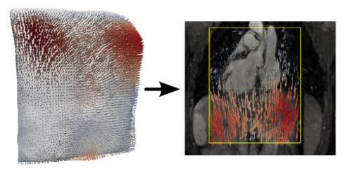
Respiratory motion is a challenge for image-guided procedures concerning the thorax or abdomen such as radiation therapy or cardiac interventions. A common approach to motion compensation is predicting internal target displacements from an external surrogate signal. Recent methods employ real-time range imaging to generate high-dimensional breathing surrogates. However, they e.g. rely on heuristic surface partitioning strategies or do not predict internal movement. We propose a statistical framework utilizing dimensionality reduction and multilinear regression to predict full internal deformation fields from dense external surface motion. In our experiments conducted on synthetic 4-D phantom and 6 volunteer data sets, we achieve a maximum prediction error of 0.42 mm given a median magnitude of the ground truth deformation field of 3.91 mm.
Articles in Conference ProceedingsProceedings of the 28th International Congress and Exhibition (Computer Assisted Radiology and Surgery (CARS 2014)), Fukuoka, Japan, 25.-28.06.2014, pp. 33-34, 2014 (BiBTeX, Who cited this?)
Jens Wetzl, Oliver Taubmann, Sven Haase, Thomas Köhler, Martin Kraus, Joachim Hornegger
-
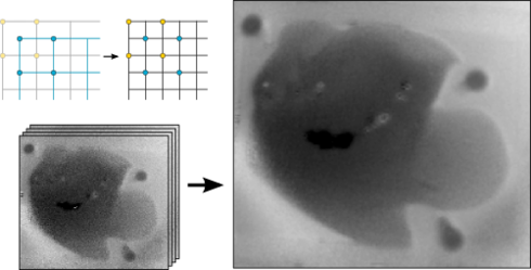
In the field of image-guided surgery, Time-of-Flight (ToF) sensors are of interest due to their fast acquisition of 3-D surfaces. However, the poor signal-to-noise ratio and low spatial resolution of today's ToF sensors require preprocessing of the acquired range data. Super-resolution is a technique for image restoration and resolution enhancement by utilizing information from successive raw frames of an image sequence. We propose a super-resolution framework using the graphics processing unit. Our framework enables interactive frame rates, computing an upsampled image from 10 noisy frames of 200×200 px with an upsampling factor of 2 in 109 ms. The root-mean-square error of the super-resolved surface with respect to ground truth data is improved by more than 20% relative to a single raw frame.
In the course of this project, we developed a
library for non-linear optimization using L-BFGS (a Quasi-Newton method) on the GPU using CUDA. The super-resolution framework that builds on it is also freely available.Articles in Conference ProceedingsBildverarbeitung für die Medizin, Heidelberg, 04.03, pp. 21-26, 2013 (BiBTeX, Who cited this?)