Dr.-Ing. Martin Berger
Alumnus of the Pattern Recognition Lab of the Friedrich-Alexander-Universität Erlangen-Nürnberg
Projects
-
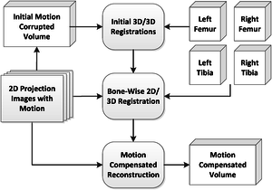
 Over the last decade, increased effort has been made to acquire three dimensional images of knee joints under weight-bearing condition. Cone-beam CT systems are popular because of their high flexibility with respect to patient position and scan trajectory. However, scans in a standing or squatting patient position are affected by involuntary patient motion during the acquisition, which results in streaking and blurring artifacts in the reconstructed volumes. Previous work suggested the use of fiducial markers to estimate and compensate for motion artifacts. However, marker placement on the skin might not accurately reflect the motion at the center of the joint. In this work, we propose a marker-free motion compensation method that is based on 2D/3D rigid registrations of individual projection images to a segmentation of the bones from a prior, motion-free scan. The estimated motion of the individual bones is then combined to a global motion field to allow for a motion compensated reconstruction. Qualitative and quantitative results show substantial improvement compared to uncorrected images. Incorporating smoothness constraints into the estimated motion parameters during the registration further improved the results.Journal ArticlesMedical Physics, vol. 43, no. 3, pp. 1235-1248, 2016 (BiBTeX, Who cited this?)Articles in Conference ProceedingsIFMBE Proceedings (World Congress on Medical Physics and Biomedical Engineering), Toronto, Canada, June 7-12, 2015, pp. 54-57, 2015, ISBN 978-3-319-19387-8 (BiBTeX, Who cited this?)
-
![Sinograms (top row) and their logarithmically scaled spectra (bottom row) for the noise-free projections. From left to right: motion corrupted, motion corrected and the motion free reference. Visualization windows are [1.0, 5.5] for the sinograms and [1.5, 5.0] for the log-spectra.](https://www5.cs.fau.de/fileadmin/_processed_/csm_sinogramsFFT_ee3a6a3745.jpg) In computed tomography involuntary patient motion can lead to a severe degradation of image quality. Most of the motion estimation methods rely on additional information, such as fiducial markers or an ECG signal. In contrast, data driven motion estimation exists which aims to estimate the motion directly from the acquired projections. This is typically achieved by the optimization of an error metric that is either defined in the image or in the projection domain. In this work, we present a novel data driven error function for motion compensation in fan-beam CT and its combination with a simple motion compensation scheme. The new method operates entirely in the fourier domain of the sinogram, by enforcing zero energy regions of the spectrum. Qualitative and quantitative results show that the proposed method is able to remove most of the motion artifacts, yielding a relative root mean square error of 7.09% compared to 20.35% for the motion corrupted reconstruction.Articles in Conference ProceedingsProceedings of the third international conference on image formation in x-ray computed tomography (The third international conference on image formation in x-ray computed tomography), Salt Lake City, UT, USA, 22-25.06.2014, pp. 203-207, 2014 (BiBTeX, Who cited this?)Proceedings of the third international conference on image formation in x-ray computed tomography (The third international conference on image formation in x-ray computed tomography), Salt Lake City, UT, USA, 22-25.06.2014, pp. 329-332, 2014 (BiBTeX, Who cited this?)
In computed tomography involuntary patient motion can lead to a severe degradation of image quality. Most of the motion estimation methods rely on additional information, such as fiducial markers or an ECG signal. In contrast, data driven motion estimation exists which aims to estimate the motion directly from the acquired projections. This is typically achieved by the optimization of an error metric that is either defined in the image or in the projection domain. In this work, we present a novel data driven error function for motion compensation in fan-beam CT and its combination with a simple motion compensation scheme. The new method operates entirely in the fourier domain of the sinogram, by enforcing zero energy regions of the spectrum. Qualitative and quantitative results show that the proposed method is able to remove most of the motion artifacts, yielding a relative root mean square error of 7.09% compared to 20.35% for the motion corrupted reconstruction.Articles in Conference ProceedingsProceedings of the third international conference on image formation in x-ray computed tomography (The third international conference on image formation in x-ray computed tomography), Salt Lake City, UT, USA, 22-25.06.2014, pp. 203-207, 2014 (BiBTeX, Who cited this?)Proceedings of the third international conference on image formation in x-ray computed tomography (The third international conference on image formation in x-ray computed tomography), Salt Lake City, UT, USA, 22-25.06.2014, pp. 329-332, 2014 (BiBTeX, Who cited this?)
-
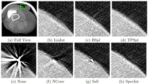
 Fiducial markers are frequently used in X-ray imaging to obtain accurate point correspondences for further processing. These markers typically cause metal artifacts, decreasing image quality of the subsequent reconstruction and are therefore often removed from the projection data. The placement of such markers is usually done on a surface, separating two materials, e.g. skin and air. Hence, a correct restoration of the occluded area is difficult. In this work six state-of-the-art interpolation techniques for the removal of high-density fiducial markers from cone-beam CT projection data are compared. We conducted a qualitative and quantitative evaluation for the removal of such markers and the ability to reconstruct the adjoining edge. Results indicate that an iterative spectral deconvolution is best suited for this application, showing promising results in terms of edge, as well as noise restoration.Articles in Conference ProceedingsBildverarbeitung für die Medizin 2014, Aachen, 16.3-18.3, pp. 168-173, 2014 (BiBTeX, Who cited this?)
-
Osteoarthritis (OA) is the leading cause of functional decline and disability in aging populations. Yet, the causes and progression of OA, particularly in the early stages, remain poorly understood. Current OA imaging measures are insensitive to early changes, or are logistically challenging and limited by expense and long scan times.
The comprehensive goal of this project is to develop novel weight-bearing computed tomography (CT) imaging to support an imaging biomarker “stress test” of the knee joint. This test will then reveal abnormal tissue biomechanics and kinematics that put a knee at high risk for OA, by measuring the articular cartilage deformation between different weight-bearing conditions.
The standard application area for C-arm CT is in the interventional suite, where it usually acquires images using a vertical trajectory around the patient. For this project we opted for a C-arm device that can be almost arbitrarily positioned in space. This allows for a horizontal trajectory which is necessary to acquire images whilst the patient is standing upright or in squatting position.
From a medical image reconstruction point of view, there exist two main challenges. First, the limited detector size of C-arm CTs leads to difficulties in acquiring images from two adjoining knees in their natural position. Therefore, one goal is to explore so called large volume techniques which extend the field of view by adjusting the imaging trajectory to the objects shape. Another goal of this project will be to investigate methods for motion correction, as the standing position results in increased patient motion during image acquisition and therefore to increased motion artefacts. Multiple methods are conceivable that give additional information about patient motion. For example metallic markers that encode the object’s position in the different detector images. Also external range imaging cameras could be used to keep track of the motion during a scan. Both methods would then allow the registration of different detector images, to allow for a correct reconstruction of the volume of interest.
This volume is then used to compute the necessary metrics of the knee joint, which can be interpreted as a biomarker for evaluating the risk of osteoarthritis.
This project is in collaboration with the Department of Radiology, Stanford University, Stanford, CA, USA.
I am a member of the research training group for heterogeneous imaging systems. For mor information click ![]() here.
here.

 +49 9131 85 27891
 +49 9131 85 27270

