
Contact


Address
Chair of Computer Science 5 (Pattern Recognition)
Germany
Powered by


Dr.-Ing. Marcus Prümmer
Alumnus of the Pattern Recognition Lab of the Friedrich-Alexander-Universität Erlangen-Nürnberg
Medical Image Reconstruction Group
Our research group is working on a wide range of topics in Medical Image Processing with the focus on tomographic reconstruction. Most of the topics are related to interventional applications using C-arm CT.

Foot Kinematics
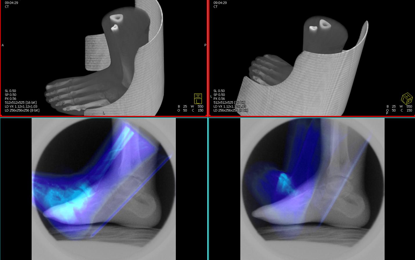
In this project foot kinematics is studied using fluoroscopy. A 3-D image of the foot is registered to a temporal sequence of fluoroscopic images. A robust and fast rigid 3-D/2-D registration method is developed and integrated into a kinematics analysis tool for bone structures. The bones in the 3-D image are segmented and registered to the 2-D projection image. The images shows an overlay of the projected foot to the fluoroscopic image (colored in blue). The foot in the image is not registered and shows a typical start position of an uncalibrated scenario.
Cooperation partners:
Non-rigid 3D-2D Registration
Motivation
In previous projects a 3D-3D registration method was performed for heart motion estimation. The motion estimation is generally subject specific and thus data driven.
One requirement to this approach is having accurate initial reconstructions to be aligned. Unfortunately the image quality of these initial reconstructions depends on the observed motion after ECG-gating. This may result depending on the scanned subject in several unfavorable blurred volume samples. Furthermore two motion blurred volumes are non-rigidly aligned, where aligned artifacts may hoke up the real motion vector field that encodes the heart motion. This motivates to come up with an alternative approach for a heart motion estimation.
The 3-D motion vector field is computed via the alignment of only one initially reconstructed volume to a series of temporal resolved (ECG-gated) projection images. The volume is reconstructed using ECG-gated Feldkamp reconstruction. This volume is warped such that its forward projections match a series of acquired projection images. The projection image series is ECG-gated as well, however this series can also be a subset of an ECG-gated short-scan set. Thus the temporal resolution can be more
focused to the cost of sparser reference projection data.
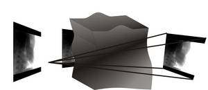
Fig. 1. Principle technique of 3-D/2-D motion estimation.
In this project a mono- and multi-modal 3D-2D non-rigid registration method is derived. The methods are based on variational calculus. Specifically the distance measures between the 3-D volume and the 2-D projection data is derived.
The objectives for the non-rigid 3-D/2-D alignment can be summarized as follows:
1. Registration output is a 3-D motion vector field (MVF).
2. Additional smoothness constraint for the MVF via spatial regularization.
3. Energy function is optimized in the space of the observed x-ray images (projection space).
4. Gradient required for a gradient-descent based optimization is computed in 3-D.
5. One initially reconstructed volume is required.
6. Sparse number (e.g. about 40 or even less) of reference projections is used to improve temporal focus.
Results

Fig. 2. Non-rigid 3-D/2-D Registration using the NCAT Phantom. From left to right:
ground truth, before alignment, aligned and first component of the computed MVF. Number of used 2-D reference projections was 13.
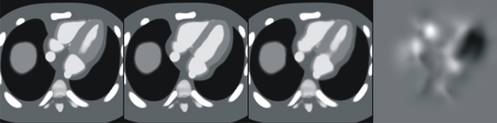
Fig. 3. Non-rigid 3-D/2-D Registration using NCAT Phantom. From left to right:
ground truth, before alignment, aligned and first component of the computed MVF. Number of used 2-D reference projections was 4.
Conclusions
The results show that using about 13 reference projections (Fig. 2.) the computed MVF represents the anatomical structure of the moving heart phantom. Bright values in the right image in Fig. 2. denote strong motion. With decreasing number of projections (compare to Fig. 3) the resolution drops significantly as shown in Fig. 3 using only 4 projections. The sparsity of four projections has to be compensated by a stronger spatial smoothness regularization of the computed MVF to still provide a stable registration. This results in a smoother MVF with a decreased spatial resolution. The experiments show that using a sparse number of time resolved (ECG-gated) reference projections, the motion can be computed between the cardiac phase of the initially reconstructed volume and the cardiac phase of the reference projections with an acceptable quality.
Cooperation with the Institute of Sport Science and Sport
 |
Parabolic flight campaign
13th DLR campaign
3 - 13 February 2009
Technical assistance and subject of an experiment for a parabolic flight campaign aboard the A300 ZERO-G.
Investigation of intermuscular coordination regarding central nervous and peripheral nervous system by means of a defined SmartCranks cycle ergometer exercise during different gravity, crank, pedaling power output and pedaling rate conditions.
Further information can be found at  www.pfm.sport.uni-erlangen.de .
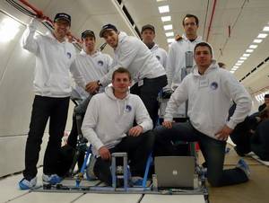
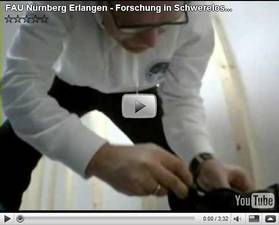
The mission crew on board of the A300 ZERO-G. Click the trailer image to watch on YouTube.
Dynamic Algebraic Reconstruction
An alternative approach to analytical reconstruction methods
is the algebraic reconstruction technique (ART). Generally spoken, the idea of algebraic
reconstruction is to define a system matrix that again defines the sampling dependency
between lattice points of the image and rays that define the spatial path during the forward projection. Thus the system matrix defines the scan geometry like parallel-, fan- or cone-beam. Given this matrix and the acquired projection data one can mathematically model the reconstruction problem as a linear equation system that needs to be solved to reconstruct the image again from its projections. We point out that the creation of the system matrix is one of the most important issues in ART.
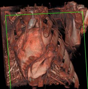
In this work we introduce a motion model of a moving object in either to the object grid, where the density values are reconstructed or to the projection geometry. For a static object grid and projection geometry, the system matrix is generally sparse, but has a regular structure. In this project we consider the creation of this system matrix and investigate the structure. Furthermore we introduce different motion models into the projector and investigate the resulting structure of a system matrix containing the dynamic geometry due to the object motion. The resulting matrix of a combined forward- and backprojection provides the point-spread-function (PSF) for each lattice point that is reconstructed. In the special case, when the PSF for each lattice point is the same, a fast filtered backprojection approach can be performed. We investigate the PSF resulting from the discretized ART system matrix for different motion models to develop a good understanding of the impact of a specific motion model to the shift-invariance or shift-variance of a system. An example of a resulting matrix of a combined forward and backprojection using a non-rigid motion model of an animal model is shown in the figure.

The image shows the resulting matrix of a combined forward- and backprojection operation using a non-rigid motion model that has been derived from an animal model.
The blocks are not Toeplitz-Block-Toeplitz anymore, as a result of a shift-variant PSF.
Cardiac C-arm CT: 4D Heart Motion Modeling and Evaluation Study
Cardiac C-arm CT is a an imaging technique under development that combines 3D cardiac image acquisition and real-time fluoroscopy on the same system. The combination (hybrid imaging system) combines the advantages of cardiac CT that is already used in 3D/4D imaging of the heart and C-arm systems that are commonly used during interventions because of their real-time projection mode, and high spatial resolution for 2D imaging. For cardiac CT imaging an ECG-gated angiographic X-ray image sequence of a specific heart phase is required to reconstruct a high quality 3D heart image. Retrospective ECG gating is an established technique used in cardiac CT systems to provide the reconstruction algorithm with X-ray images of the heart in the phase corresponding to the phase that is reconstructed. This requires a high temporal resolution and places therefore stringent requirements on the hardware for C-arm CT which are by current hardware implementations not fulfilled. In order to approach the temporal resolution of clinical CT with C-arm CT hardware, we are investigating novel image processing algorithms for non-parametric heart motion modeling to correct the data used in the image reconstruction process to reduce motion blurring. 3D ultrasound systems are used for evaluation of the reliability of the different non-parametric heart motion models based on real cardiac data. Such systems allow the quantification of the true heart motion.
Cardiac C-arm CT: Efficient Motion Correction for 4D-FBP
Cooperation Partners:
Department of Radiology, Stanford University.
Siemens AG, Medical Solutions, Forchheim, Germany.
Cardiac C-arm CT is a promising technique that enables 3D cardiac image acquisition and real-time fluoroscopy on the same system. The goal is to bring 3D imaging to the interventional suite for improved therapy planning, guiding, and monitoring. For the reconstruction of 3D cardiac image data, a complete set of projections from a specific heart phase is required. One approach to reduce motion blurring caused by the beating heart is to acquire multiple sweeps using the C-arm and retrospectively select the projections (RECG) that are closest to the desired cardiac phase [1]. In order to further improve the temporal resolution, novel image processing algorithms that utilize retrospective motion correction were investigated in this project. The main focus of this work is to extend the well established FDK algorithm to incorporate motion correction during the back-projection step using a pre-computed motion field. In our experiments we investigated the following two scenarios: (i) Can the image quality from a single sweep be improved given a known motion field? (ii) Can improved image quality be achieved using a lower number of sweeps in combination with motion correction?
Cardiac C-arm CT protocols are, among other things, constrained by the rotation speed and frame rate of the C-arm and detector, the breath hold duration, overall injected dose of contrast agent, scan synchronization with the ECG and X-ray dose. First empirical studies show that protocols of four sweeps are reasonable for images acquired in diastoly, but temporal resolution can still be improved because of heart rate variations during the scan. We have shown that increased temporal resolution can be achieved using a first order motion estimation via 3D-3D non-rigid registration applied on a pure RECG reconstructed time series, that is blurred and has motion artifact, with correction applied in an extended FDK (FDK-4D) algorithm. For scenario (i) we showed that assuming a 4D motion field is given, the FDK-4D algorithm is able to decrease motion blurring significantly using only one single sweep for the reconstruction. For scenario (ii) we showed that even for an insufficient ECG synchronization using three and four sweeps, the reconstructed images of the diastolic and systolic phase are less blurred using FDK-4D compared to pure RECG. In conclusion, increasing temporal resolution using an estimated 4D motion field in the FDK-4D algorithm can decrease motion blurring substantially.
This work was headed by Rebecca Fahrig and supported by Siemens AG, Medical Solutions, NIH grant R01 EB 003524 and by the Lucas Foundation.
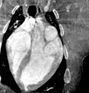
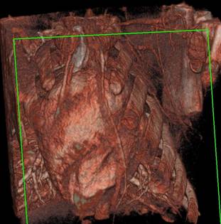
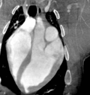
The top row shows a retrospectively ECG-gated Feldkamp reconstruction using 191 projection images. The left column shows a MPR (windowing width=760 center=99) of a long axis view from a pigs heart in a systolic cardiac phase and the right column the corresponding volume rendered image (windowing width=426, center=322). In the bottom row the corresponding MPR and volume rendered image is shown that was reconstructed using motion correction. For this motion corrected reconstruction all 1146 acquired projection images of a 6x4s multi-sweep C-arm run were taken into account during reconstruction.
Cardiac C-arm CT: 4D Non-Model based Heart Motion Estimation and its Application
In principal, to reconstruct a 3D-image of the beating heart at a particular cardiac phase, a complete set of X-ray projection data representing that phase isrequired. One approximate approach is the retrospectively ECG-gated FDK reconstruction (RG-FDK). From the acquireddata set of Ns multiple C-arm sweeps, those projection images which are acquired closest in time to the desired cardiacphase are retrospectively selected. However, this approach uses only 1/Ns of the obtained data. Our goal is to utilize data fromother cardiac phases as well. In order to minimize blurring and motion artifacts, cardiac motion has to be compensated for,which can be achieved using a temporally dependent spatial 3D warping of the filtered-backprojections. In this work weinvestigate the computation of the 4D heart motion based on prior reconstructions of several cardiac phases using RG-FDK. A 4D motion estimation framework is presented using standard fast non-rigid registration. A smooth 4D motion vectorfield (MVF) represents the relative deformation compared to a reference cardiac phase. A 4D deformation regridding byadaptive supersampling allows selecting any reference phase independently of the set of phases used in the RG-FDK fora motion corrected reconstruction. Initial promising results from in vivo experiments are shown. The subjects individual4D cardiac MVF could be computed from only three RG-FDK image volumes. In addition, all acquired projection datawere motion corrected and subsequently used for image reconstruction to improve the signal-to-noise ratio compared to RG-FDK.
Cardiac C-Arm CT: Image-based Gating
Applying the concept of electrocardiogram gating (ECG) during the acquisition of multiple,serial, backward and forward, ECG-triggered rotational acquisitions using a C-arm system allows the 3D+t reconstructionof the heart. The process of retrospective gating is a crucial component of 3-D reconstruction. The gold-standard is still given by the ECG signal. However, an alternative gating method, based on the acquired projection data is required. Our goal is to provide an image-based gating method without ECG.
Further Projects
- SNR enhanced reconstruction: we derive and compare weighting schemes that combine motion corrected reconstructions.
- Comparison between different gating methods: relative cardiac phase vs. absolute time.
- Development of an ultrasound evaluation framework to investigate motion estimation accuracy.