Dr.-Ing. Jan Paulus
Alumnus of the Pattern Recognition Lab of the Friedrich-Alexander-Universität Erlangen-Nürnberg
Automatic Quality Assessment of Diffusion Weighted Images
|
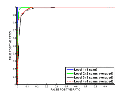
Diseases like glaucoma affect the visual pathway in the brain. Diffusion Tensor Imaging is the only imaging modality to identify the related fibers (see The following local and global image statistics are combined to perform quality assessment. (i) Structural recognizability is implemented by an unsupervised clustering, that detects grey/white matter, the background and a last cluster representing the remaining regions in the brain (e.g. cerebrospinal fluid and air). It calculates the cluster sizes and inter-cluster-contrasts locally. (ii) A sharpness metric measures the local structural differentiation by counting strong edges and calculating their average strength. (iii) The Haralick features entropy, energy and contrast judge the global sharpness, homogeneity and contrast. The three feature groups are combined and directly used for quality level classification. Diffusion weighted images of 10 subjects were scanned along 20 gradient directions. Our gold standard consisted of four quality levels (1-4) starting with the original images (level 1) and improving by averaging an increasing number of images (2-4). A cross-validation strategy was performed on the whole data set. A probabilistic Support Vector Machine was used for classifying the four quality levels. The method reaches an area under the ROC curve of above 97.0% for correctly classifying each quality level. It shows the highest performance for the lowest quality level. We developed a method for automatically classifying different quality levels in diffusion weighted images independently from gradient directions. It reaches the highest performance in classifying lowest quality. The proposed automated method is able to measure diffusion weighted image quality successfully and to produce reliable results. |
|
2010 (1 Publications, Talks and Patents)
Talks
Automatic Quality Assessment of Diffusion Weighted Images
at ARVO/International Society for Imaging in the Eye (ISIE) (Annual Meeting) in Fort Lauderdale, FL, USA (01.05.)
(BiBTeX, Who cited this?) ![]() Poster
Poster


