Contact


Address
Chair of Computer Science 5 (Pattern Recognition)
Martensstrasse 3
91058 Erlangen
Germany
Powered by


Dr.-Ing. Arne Militzer
Alumnus of the Pattern Recognition Lab of the Friedrich-Alexander-Universität Erlangen-Nürnberg
Virtual Planning of Liver Interventions
 |
Primary liver tumors are among the most frequent primary malignant tumors. Secondary liver tumors are even more common, amounting for about 90% of all liver lesions[1]. Especially colon cancer patients have a very high risk of developing liver metastases at some point [2]. However, so far computer assistance for physicians during diagnosis and surgical treatment of liver tumors is usually very basic.
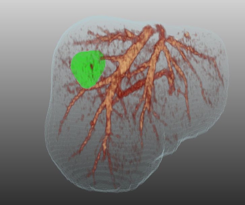
This project aims at overcoming this deficit by providing an intervention planning system that, given a set of CT images, automatically detects and segments liver lesions, characterizes them, and analyzes their position relative to important anatomical structures like vessels or liver segments. This information can then not only be visualized in 3-D (Fig. 1) for surgeons to get a better orientation. It is also used to simulate interventions including side eff ects such as liver regions being cut off their blood supply or drainage due to damaged vascular structures. Additionally, with the segmentation available, tumors that have not been resected can be monitored over a longer period of time in order to allow an assessment of tumor growth or shrinkage and thus the success of the treatment.
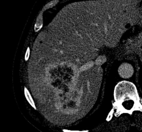 |
A major contribution of this work will be the automatic detection and segmentation of liver lesions. In particular, we want to focus on inhomogenous lesions and lesions showing a strong enhancement along the tumor boundary (Fig. 2). Current semi-automatic segmentation methods, which are often based on thresholding or region growing techniques, usually succeed for well-shaped, homogenous tumors, but fail for the mentioned more difficult cases. Therefore, machine learning techniques will be applied to robustly detect and segment these tumors in images. To achieve this robustness and reliability, information from various sources, such as CT images of
different phases of contrast enhancement in the liver, will be incorporated into the process.
References
1. Layer, G; Gallkowski, U.: Lebertumoren. Chap.5 of Radiologische Diagnostik in der Onkologie: Band 2 Gastrointestinum, Urogenitaltrakt, Retroperitoneum. Springer Verlag Berlin Heidelberg, 2008, ISBN 978-3-540-29318-7
2. Layer, G.; Kopp, A. ; Müller-Schimpe, M.: Kolorektales Karzinom. Chap.4 of Ra diologische Diagnostik in der Onkologie: Band 2 Gastrointestinum, Urogenitaltrakt, Retroperitoneum. Springer Verlag Berlin Heidelberg, 2008, ISBN 978-3-540-29318-7
This project is funded by Siemens AG Healthcare, Computed Tomography, Forchheim, Germany.