Dept. of Computer Sc. » Pattern Recognition » Our Team » Mayer, Markus » Automated Glaucoma Classification using OCT Images

Dipl.-Inf. Markus Mayer
Alumnus of the Pattern Recognition Lab of the Friedrich-Alexander-Universität Erlangen-Nürnberg
The vision of my work is to develop image processing methods that provide new tools for ophthalmologists to ease the detection of glaucoma and related eye diseases.
Automated Glaucoma Classification Using OCT Images
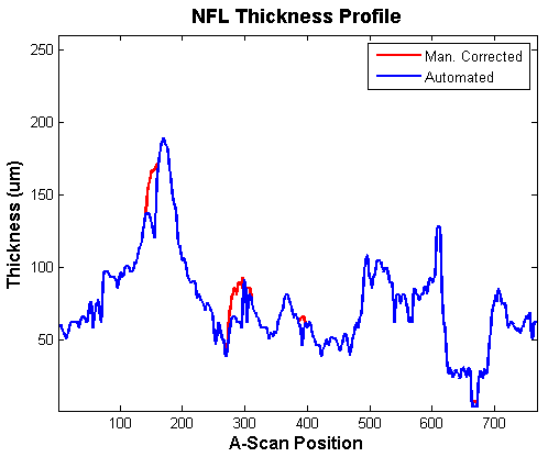
The glaucoma disease is the loss of supporting tissue and nerve fibers in the papilla area. OCT is capable to image this region in depth, and thus give an in-vivo impression of the retinal nerve fiber layer thickness. Using thickness profiles generated out of our  Automated Retinal Layer Segmentation we try to automatically discriminate between normals and glaucoma patients. The goal is to develop an OCT-based glaucoma indicator comparable to the GPS included in the HRT imaging device and the
  Glaucoma Risk Index (GRI) for color fundus photographs invented at our lab.
 |
Preliminary work on this project is published in the following form:
- Markus A. Mayer, Joachim Hornegger, Christian Y. Mardin, Friedrich E. Kruse, Ralf. P. Tornow: "Automated Glaucoma Classification Using Nerve Fiber Layer Segmentations On Circular Spectral Domain OCT B-Scans", The Association for Research in Vision and Ophthalmology, Inc. (ARVO) (Annual Meeting) in Fort Lauderdale, Florida, USA, 2009. Poster Download


