Contact

+49 9131 85 28977

+49 9131 85 27270
Address
Universität Erlangen-Nürnberg
Chair of Computer Science 5 (Pattern Recognition)
Martensstraße 3
91058 Erlangen
Germany
Powered by

|
Thomas Köhler M. Sc.
Alumnus of the Pattern Recognition Lab of the Friedrich-Alexander-Universität Erlangen-Nürnberg
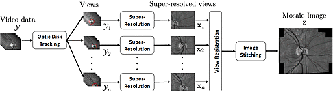
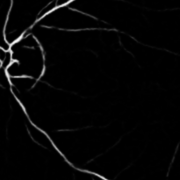
Super-Resolved Retinal Image Mosaicing
Thomas Köhler, Axel Heinrich, Andreas Maier, Joachim Hornegger, Ralf P. Tornow
-
The acquisition of high-resolution retinal fundus images with a large field of view (FOV) is challenging due to technological, physiological and economic reasons. This paper proposes a fully automatic framework to reconstruct retinal images of high spatial resolution and increased FOV from multiple low-resolution images captured with non-mydriatic, mobile and video-capable but low-cost cameras. Within the scope of one examination, we scan different regions on the retina by exploiting eye motion conducted by a patient guidance. Appropriate views for our mosaicing method are selected based on optic disk tracking to trace eye movements. For each view, one super-resolved image is reconstructed by fusion of multiple video frames. Finally, all super-resolved views are registered to a common reference using a novel polynomial registration scheme and combined by means of image mosaicing. We evaluated our framework for a mobile and low-cost video fundus camera. In our experiments, we reconstructed retinal images of up to 30° FOV from 10 complementary views of 15° FOV. An evaluation of the mosaics by human experts as well as a quantitative comparison to conventional color fundus images encourage the clinical usability of our framework.
Supplementary material:
 Here, we provide an example video to demonstrate super-resolved mosaicing on example datasets of two human subjects. Please refer to our paper for details of the experimental evaluation. Here, we provide an example video to demonstrate super-resolved mosaicing on example datasets of two human subjects. Please refer to our paper for details of the experimental evaluation.- The presentation slides for this work are available
 here. here.
2016 IEEE 13th International Symposium on Biomedical Imaging (International Symposium on Biomedical Imaging (ISBI))), Prague, Czech Republic, 2016, pp. 1063-1067, 2016 (BiBTeX, Who cited this?)
Multi-Frame Super-Resolution with Quality Self-Assessment for Retinal Fundus Videos
Thomas Köhler, Alexander Brost, Katja Mogalle, Qianyi Zhang, Christiane Köhler, Georg Michelson, Joachim Hornegger, Ralf P. Tornow
-
This paper proposes a novel super-resolution framework to reconstruct high-resolution fundus images from multiple low-resolution video frames in retinal fundus imaging. Natural eye movements during an examination are used as a cue for super-resolution in a robust maximum a-posteriori scheme. In order to compensate heterogeneous illumination on the fundus, we integrate retrospective illumination correction for photometric registration to the underlying imaging model. Our method utilizes quality self-assessment to provide objective quality scores for reconstructed images as well as to select regularization parameters automatically. In our evaluation on real data acquired from six human subjects with a low-cost video camera, the proposed method achieved considerable enhancements of low-resolution frames and improved noise and sharpness characteristics by 74%. In terms of image analysis, we demonstrate the importance of our method for the improvement of automatic blood vessel segmentation as an example application, where the sensitivity was increased by 13% using super-resolution reconstruction.
Supplementary material: - The poster for this work is available here.
Here, we provide an example video for a comparison of low-resolution video data and super-resolved fundus images (super-resolution applied to 8 successive frames with magnification factor 2, input frame rate: 12.5 Hz). Please see our paper for further details on image acquisition. Source code: MATLAB source code for our method is available here - Our software includes the latest version of our super-resolution toolbox (including demo scripts and test data).
- This software requires the Image Alignment Toolbox (current version: 0.9.1) for eye motion estimation.
Medical Image Computing and Computer-Assisted Intervention -- MICCAI 2014 (International Conference on Medical Image Computing and Computer-Assisted Intervention), Cambridge, MA, 2014, pp. 650-657, 2014 (BiBTeX, Who cited this?)
Computer Aided Diagnostics and Pattern Recognition: Automated Glaucoma Detection
Thomas Köhler, Rüdiger Bock, Joachim Hornegger, Georg Michelson
-
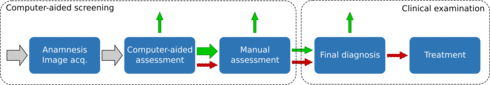
Glaucoma is one of the major causes for blindness with a high rate of unreported cases. To reduce this number, screening programs are performed. However, these are characterized by a high workload for manual and cost-intensive assessment. Computer-aided diagnostics (CAD) to perform an automated pre-exclusion of normals might help to improve program's efficiency.
This chapter reviews and discusses recent advances in the development of pattern recognition algorithms for automated glaucoma detection based on structural retinal image data. Two main methodologies for glaucoma detection are introduced: (i) structure-driven approaches that mainly rely on the automated extraction of specific medically relevant indicators, while (ii) data-driven techniques perform a generic machine-learning approach on entire image data blobs. Both approaches show a reasonable and comparable performance although they rely on different basic assumptions. A combination of these might further improve CAD for a more efficient and cost-sensitive workflow as a major proportion of normals will be excluded from unnecessary detailed investigations.
Teleophthalmology in Preventive Medicine, Springer Berlin Heidelberg Berlin, 2015, 93-104 (BiBTeX, Who cited this?)
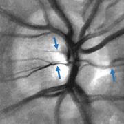
Geometry-Based Optic Disk Tracking in Retinal Fundus Videos
Anja Kürten, Thomas Köhler, Attila Budai, Ralf-Peter Tornow, Georg Michelson, Joachim Hornegger
-
Fundus video cameras enable the acquisition of image sequences to analyze fast temporal changes on the human retina in a non-invasive manner. In this work, we propose a tracking-by-detection scheme for the optic disk to capture the human eye motion on-line during examination. Our approach exploits the elliptical shape of the optic disk. Therefore, we employ the fast radial symmetry transform for an efficient estimation of the disk center point in successive frames. Large eye movements due to saccades, motion of the head or blinking are detected automatically by a correlation analysis to guide the tracking procedure. In our experiments on real video data acquired by a low-cost video camera, the proposed method yields a hit rate of 98 % with a normalized median accuracy of 4 % of the disk diameter. The achieved frame rate of 28 frames per second enables a real-time application of our approach.
Automatic No-Reference Quality Assessment for Retinal Fundus Images Using Vessel Segmentation
Thomas Köhler, Attila Budai, Martin Kraus, Jan Odstrcilik, Georg Michelson, Joachim Hornegger
-
Fundus imaging is the most commonly used modality to collect information about the human eye background. Objective and quantitative assessment of quality for the acquired images is essential for manual, computer-aided and fully automatic diagnosis. In this paper, we present a no-reference quality metric to quantify image noise and blur and its application to fundus image quality assessment. The proposed metric takes the vessel tree visible on the retina as guidance to determine an image quality score. In our experiments, the performance of this approach is demonstrated by correlation analysis with the established full-reference metrics peak-signal-to-noise ratio (PSNR) and structural similarity (SSIM). We found a Spearman rank correlation for PSNR and SSIM of 0.89 and 0.91. For real data, our metric correlates reasonable to a human observer, indicating high agreement to human visual perception.
Supplementary material: - The presentation slides for this work are available here.
2013 26th IEEE International Symposium on Computer-Based Medical Systems (CBMS) (International Symposium on Computer-Based Medical Systems), Porto, Portugal, 2013, pp. 95-100, 2013 (BiBTeX, Who cited this?)
Quality-Guided Image Denoising for Low-Cost Fundus Imaging
Thomas Köhler, Joachim Hornegger, Markus Mayer, Georg Michelson
-
The restoration of noisy images is an essential pre-processing step in many medical applications to ensure sufficient quality for diagnoses. In this paper we present a new quality guided approach for denoising of eye fundus images that suffer from high noise levels. The denoising is based on image sequences and an adaptive frame averaging approach. The novelty of the method is that it takes an objective image quality criteria to assess the different frames and tries to maximize the quality of the resulting image. It can be implemented in an incremental manner which allows real-time denoising. We evaluated our approach on real image sequences captured by a low-cost fundus camera and obtained competitive results to a state-of-the-art method in terms of signal-to-noise ratio whereas our method performs denoising about four times faster.
Supplementary material:
- The presentation slides for this work are available here.
Bildverarbeitung für die Medizin 2012 (Workshop Bildverarbeitung für die Medizin), Berlin, 19.03.2012, pp. 292-297, 2012, ISBN 978-3-642-28502-8 (BiBTeX, Who cited this?)
|


 high-resolution fundus (HRF) database
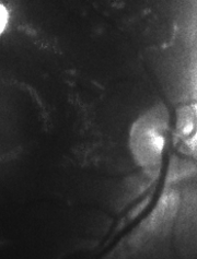