
Contact


Address
Chair of Computer Science 5 (Pattern Recognition)
Germany
Powered by


Dr.-Ing. Florian Jäger
Alumnus of the Pattern Recognition Lab of the Friedrich-Alexander-Universität Erlangen-Nürnberg
Computer-Aided-Assessment of Anomalies in the Scoliotic Spine in 3-D MRI Images
The assessment of anomalies in the scoliotic spine using Magnetic Resonance Imaging (MRI) is an essential task during the planning phase of a patient's treatment and operations. Due to the pathologic bending of the spine, this is an extremely time consuming process as an orthogonal view onto every vertebra is required. In this project work we develop a system for computer-aided assessment (CAA) of anomalies in 3-D MRI images of the spine relying on curved planar reformations (CPR). The MRI protocol used is SPACE (Siemens, Erlangen, Germany). We treat all necessary steps, from the pre-processing of the data to the visualization component. The core part of our framework is a segmentation of the spinal channel and cord. The developed segmentation method is an iterative process. In every iteration the segmentation is updated by an energy based scheme derived from Markov random field (MRF) theory. We evaluate the segmentation results on clinical relevant 3-D MRI data sets of scoliosis patients. The data sets can be found on this page. In order to assess the quality of the segmentation we use the angle between automatically computed planes through the vertebra and planes estimated by medical experts. In order to receive the reference planes please write an Email to Florian Jäger (jaeger (at) informatik (dot) uni-erlangen (dot) de). Our experiments show that using our method yields a mean angle difference of less than six degrees.
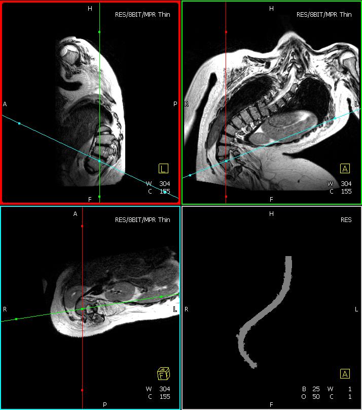
The image shows a typical segmentation result for a scoliosis patient (bottom right). From the segmentation the central axis is computed and then the CPR for the current slider position (bottom left). In the upper row a sagittal and a coronal slice through the data set is shown.
Public Clinical Relevant 3-D SPACE Images of Scoliosis Patients
We would like to know who is using our data sets. For this reason we would appreciate it if you send us the following information.
If you are using the data sets for a publication, please reference the following article:
Florian Jäger, Joachim Hornegger, Siegfried Schwab and Rolf Janka: "Computer-Aided Assessment of Anomalies in the Scoliotic Spine in 3-D MRI Images". In MICCAI, London, UK, Springer (2009), accepted for publication