
Contact


Address
Chair of Computer Science 5 (Pattern Recognition)
Germany
Powered by

Dr.-Ing. Matthias Weidler
Alumnus of the Pattern Recognition Lab of the Friedrich-Alexander-Universität Erlangen-Nürnberg
Catheter Detection and Reconstruction
Electro-physilolgy (EP) procedures are used for treatment of heart arrhythmias. They are done minimally invasive using fluoroscopic guidance. As a single X-ray image does not provide depth information, biplane systems can be used to acquire simultaneously images from two different directions. So, the physician can mentally derive the 3-D shape of the catheters. This, however, requires much experience.
Basically, a 3-D shape of a object can be computed from two views if point correspondences are known. Manual annotation of these correspondences is time consuming. Therefore, we develop approaches to automatically detect and reconstruct catheters.
-
Curvilinear objects can be reconstructed from two projection images if point correspondences are known. Possible point correspondences for a point belonging to the curve in A can be computed by intersecting its epipolar line with the curve in B. We evaluate a selection strategy which can deal with multiple (solid yellow line) or missing (solid green line) intersections. 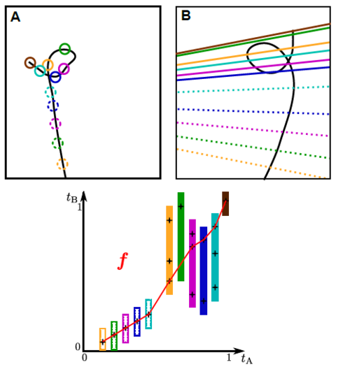
Minimally invasive interventions often involve tools of curvilinear shape like catheters and guide-wires. If the camera parameters of a fluoroscopic system or a stereoscopic endoscope are known, a 3-D reconstruction of corresponding points can be computed by triangulation. Manual identification of point correspondences is time consuming, but there exist methods that automatically select corresponding points along curvilinear structures. The focus here is on the evaluation of a recent published method for catheter reconstruction from two views. A previous evaluation of this method using clinical data yielded promising results. For that evaluation, however, no 3-D ground truth data was available such that the error could only be estimated using the forward-projection of the reconstruction. In this paper, we present a more extensive evaluation of this method based on both clinical and phantom data. For the evaluation using clinical images, 36 data sets and two different catheters were available. The mean error found when reconstructing both catheters was 0.1mm ± 0.1mm. To evaluate the error in 3-D, images of a phantom were acquired from 13 different angulations. For the phantom, A 3-D C-arm CT voxel data set of the phantom was also available. A reconstruction error was calculated by comparing the triangulated 3D reconstruction result to the 3D voxel data set. The evaluation yielded an average error of 1.2mm ± 1.2mm for the circumferential mapping catheter and 1.3mm ± 1.0mm for the ablation catheter.
Link to paper coming soon.
 View poster
Hoffmann, Matthias; Brost, Alexander; Jakob, Carolin; Bourier, Felix; Koch, Martin; Kurzidim, Klaus; Hornegger, Joachim; Strobel, Norbert
-
Detection result of the proposed single-click catheter detection. For reconstruction using corresponding points, a selection strategy is proposed for determining the best-fitting set of point correspondences. 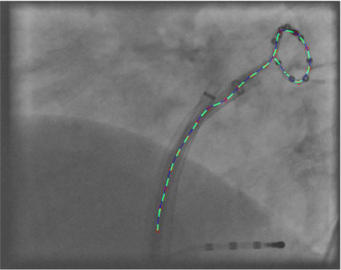
We propose novel methods for (a) detection of a catheter in fluoroscopic images and (b) reconstruction of this catheter from two views. The novelty of (a) is a reduced user interaction and a higher accuracy. It requires only a single seed point on the catheter in the fluoroscopic image. Using this starting point, possible parts of the catheter are detected using a graph search. An evaluation of the detection using 66 clinical fluoroscopic images yielded an average error of 0.7 mm ± 2.0 mm. The novelty of (b) is a better ability to deal with highly curved objects as it selects an optimal set of point correspondences from two point sequences describing the catheters in two fluoroscopic images. The selected correspondences are then used for computation of the 3-D reconstruction. The evaluation on 33 clinical biplane images yielded an average backprojection error of 0.4 mm ± 0.6 mm.
Articles in Conference ProceedingsMICCAI 2012, Part I (International Conference on Medical Image Computing and Computer Assisted Intervention), Nice, France, 01.10.2012, pp. 584-591, 2012 (BiBTeX, Who cited this?)
 View paper