
Contact


Address
Chair of Computer Science 5 (Pattern Recognition)
Martensstrasse 3
91058 Erlangen
Germany
Powered by

Dr.-Ing. Robert Grimm
Alumnus of the Pattern Recognition Lab of the Friedrich-Alexander-Universität Erlangen-Nürnberg
-
Dynamic imaging of the lung and abdomen requires a trade-off between spatial resolution, temporal resolution, and total acquisition time. Due to its robustness against motion, the clinical use of free-breathing 3D radial spoiled gradient echo (VIBE) imaging for abdominal applications has recently gained interest. It has also been shown that self-gating techniques enable the reconstruction of different respiratory phases and compensation of respiratory motion in dynamic contrast-enhanced MRI (DCE-MRI). Typically, however, separate acquisitions are required to capture the respiratory movement and dynamic perfusion. We show that a retrospective self-gating technique can be employed to extract all information from a single continuous free-breathing scan acquired with a stack-of-stars 3D VIBE sequence. This technique can be used to compute different sliding-window reconstructions that show a complete ‘virtual’ respiratory cycle with high spatial resolution, or, alternatively, a time series of the contrast-enhancement at a specified level of inspiration and with high temporal resolution.
 Related Publication:Proceedings of International Society for Magnetic Resonance in Medicine (ISMRM 20th Annual Meeting), Melbourne, Australia, May 5-11, pp. 3814, 2012 (BiBTeX, Who cited this?)
-
Recently, novel Compressed Sensing methods have been proposed that promise very high undersampling factors in the spatial as well as temporal domain. However, it is unclear if these advanced reconstruction techniques preserve the true dynamics of the contrast enhancement, which otherwise could have significant impact on the quantitative analysis of the contrast-agent kinetics. In the latter case, a noisy image that reflects the true underlying dynamics would be more favorable than a beautified image that shows less noise and undersampling artifacts but deviates from the true course of contrast enhancement. To investigate these effects, we developed an extension to the well-known analytical Shepp-Logan phantom in the temporal dimension to simulate contrast enhancement in arteries and in healthy and pathological tissue. Unlike image-based approaches, a k-space based phantom allows for accurate sampling along arbitrary trajectories. This enables assessing the influence of k-space sampling strategies as well as the evaluation of reconstruction techniques for dynamic imaging. MATLAB source code for the phantom as well as for the generation of the contrast dynamics will be made available online
 here.
here. Related Publication:Proceedings of International Society for Magnetic Resonance in Medicine (ISMRM 20th Annual Meeting), Melbourne (Australia), May 5-11, pp. 2559, 2012 (BiBTeX, Who cited this?)
-
Radial sampling of k-space allows to deduce information about the respiratory cycle directly from the data (“self-gating”).
Such signals are often biased by imperfect gradient timings, leading to an angular modulation. For golden-angle trajectories with an angular increment of 111.25°, this causes high-frequency variations. Conventional band-pass filtering reduces the fluctuations but causes loss of temporal accuracy. We propose a robust filtering scheme to suppress the angular dependency in self-gating signals extracted from golden-angle radial MRI data. The proposed filter tolerates irregular breathing and preserves temporal resolution.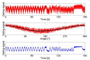 Related Publication:Proceedings of International Society for Magnetic Resonance in Medicine (ISMRM 19th Annual Meeting), Montréal, Québec, Kanada, 07.-13.05.2011, vol. 19, pp. 2677, 2011 (BiBTeX, Who cited this?)
Radial MRI
In Magnetic Resonance Imaging (MRI), image acquisition takes place in the frequency domain. For samples taken on a cartesian grid, the image is reconstructed by simply computing the 2D Fast Fourier Transform of the measurement data. With current hardware, also non-cartesian trajectories like spirals or radial spokes are feasible. Compared to cartesian trajectories, they offer several advantages such as improved robustness against motion artifacts. | 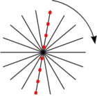 |
| To accelerate radial MRI, the number of radial spokes can be reduced. This undersampling leads to streaking artifacts in the reconstructed image: |  |
Iterative Reconstruction
MR image reconstruction can also be formulated as solving the linear equation A x = y, where x is the (unknown) image, y the observed data, and A the forward system matrix. For fully sampled cartesian data, A could simply be the matrix that applies the Discrete Fourier Transform to vector x. In general, the equation cannot be inverted directly. Instead, an iterative optimization scheme estimates the image that matches the observations best. A regularization term is incorporated to enforce sparsity of the estimated image in the Total Variation (TV) domain, or, in other words, to penalizes the streaking artifacts. With a weighting factor of λ for the TV penalty, the unconstrained optimization problem is: Minimize ||A x - y||2 + λ RTV( x ). | 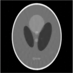 |