Friedrich-Alexander-Universität Erlangen
Lehrstuhl für Mustererkennung
Martensstraße 3
91058 Erlangen

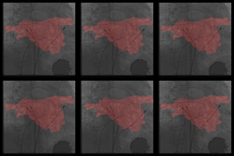
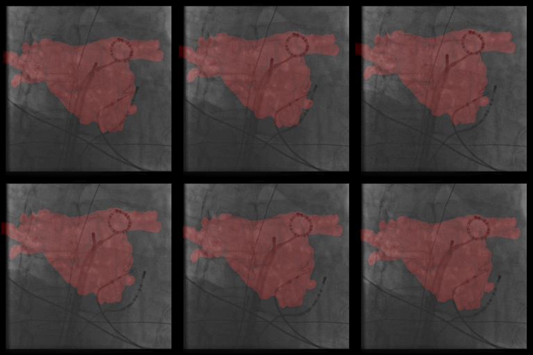
Atrial fibrillation is the most common sustained heart arrhythmia and a leading cause of stroke. The treatment by catheter ablation, performed using fluoroscopic image guidance, has become the treatment of choice. Such procedures are performed in electrophysiology labs equipped with modern C-arm X-ray systems. Fluoroscopic overlay images rendered from pre-operative volumetric data, e.g., CT, MRI or C-Arm CT, provide additional guidance for physicians during these catheter ablation. As these overlay images are compromised by cardiac and respiratory motion, we propose a method to provide motion compensation by tracking a commonly used mapping catheter.
A catheter model is generated by manual initialization. The catheter is segmented by a boosted classifier cascade. Afterwards, a model-based registration is applied to perform motion compensation. We achieve a frame rate that is higher than the clinical demands and our accuracy is as low as the thickness of the catheter.
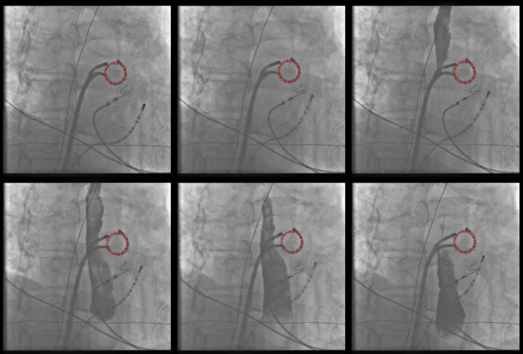
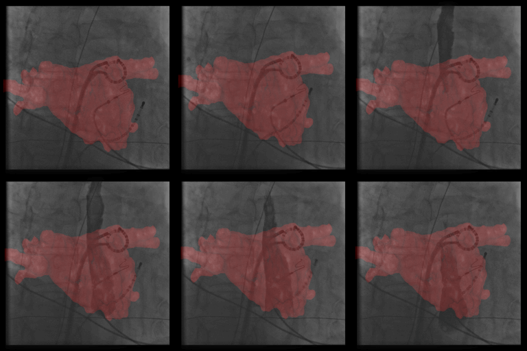
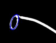
In fluoroscopic guided electrophysiology procedures, only 2-D projection images are available. To provide the physician 3-D information about the catheter positions, we reconstruct catheters from two views.
Our methods include the reconstruction of the cryo-balloon catheter and the circumferential mapping catheter.
 |
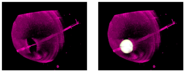 |
Cryo-balloon catheter are so-called single-shot-devices. The planning of cryo-balloon ablations for treatment of atrial fibrillation is a crucial task for a physician as he has to determine which size of the balloon catheter is required for isolation at each pulmonary vein. We Atrial Fibrillation Ablation Planning Tool (AFiT) is the first tool that provides physician a support for this planning task.
