Friedrich-Alexander-Universität Erlangen
Lehrstuhl für Mustererkennung
Martensstraße 3
91058 Erlangen

My PhD project focuses on the correction of involuntary motion during C-arm CT scans using biomechanical modeling.
Osteoarthritis (OA) is the leading cause of functional decline and disability in aging populations. Its causes and progression in the early stages remain poorly understood. The main disadvantages of current OA imaging systems are that they fail to provide information about early changes and that they require long scan times.
A novel approach to quantitatively analyze the knee joint is pursued in this project. The overarching goal is to develop a computed tomography (CT) imaging method to scan the patients in a standing/squatting and thereby weight-bearing position. This can help to further understand the mechanical stresses on the knee joint that affect cartilage and meniscus health. The scanner used in the project is a flexible C-arm CT that is able to run a horizontal trajectory.
However, scanning the patients in a standing or squatting position will lead to involuntary motion of the knees during the scan. This motion will result in artifacts in the reconstruction that reduce the diagnostic image quality. Different approaches to compensate the motion have already been investigated in previous parts of the project.
The project is in collaboration with the ![]() Department of Radiology, Stanford University, Stanford, CA, USA.
Department of Radiology, Stanford University, Stanford, CA, USA.![]()

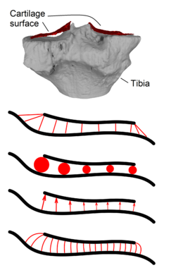
Cartilage thickness and especially its change over time is an indicator for knee cartilage health. This is especially important for Osteoarthritis (OA) patients. OA is a degenerative disease affecting bones and cartilage especially in the human knee. Thickness measurements can be performed on medical images acquired in-vivo.
Currently, there is no standard method agreed upon that defines a distance measure in articular cartilage. For this reason, we investigated the similarities and differences of methods commonly used in literature applied to the same C-arm CT data set.


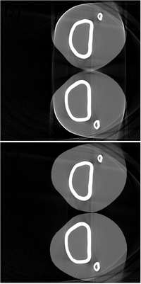
Flat-Panel C-arm Computed Tomography (CT) sufferel saturation due to the detector’s limited dynamic range. This is the case expecially when the s from pixscanned objects have different thicknesses and absorption behavior and a large dose is required.
To prevent detector saturation, a non-linear transformation of intesities is applied in the analog domain. An analog TM operator (TMO) applies a non-linear transformation in a CMOS (complementary metal-oxide semiconductor) sensor and its inverse TMO based on 14-bit digital raw data.
By this, image quality and low-contrast visibility can be improved. An application to simulated Cone-beam C-Arm CT data of two knees and a low-contrast head phantom showed qualitative as well as quantitative enhancement.
