Friedrich-Alexander-Universität Erlangen
Lehrstuhl für Mustererkennung
Martensstraße 3
91058 Erlangen

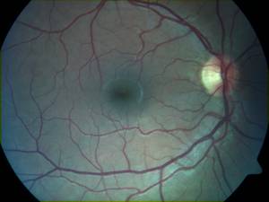
The aim of the project is to prove the usefulness of spectral filtered fundus images developed at the eye clinic in Erlangen. These RGB images are similar to the common fundus images, but none of the three channels are under- or oversaturated. Due to the colour balance these images provide more information compared to the common fundus photographs. We have to extract and show this information gain with measurements of features which were not available until now, and we have to adjust the quality measurement algorithms to check the three colour channels one by one, and give a feedback about which filter setting looks optimal for different tasks.
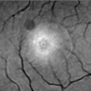
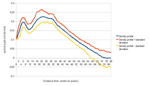
The following pictures show one of the mentioned features: the optical density of macular pigments is calculated from the absorbtion difference in the blue and green visible light domain. The semiautomatic algorithm needs a manually selected ROI which contains the macula region. In this region the algorithm detects the center of the macula, and calculates an optical density profile of the pigments.
 | Spectral filtered fundus image: |
 | Normalized absorbtion difference of blue and green channels in the selected ROI |
 | Density profile: |