Friedrich-Alexander-Universität Erlangen
Lehrstuhl für Mustererkennung
Martensstraße 3
91058 Erlangen

The aim of the project is to develop vessel segmentation and feature extraction algorithms to analyse different attributes of the vessels and the vessel tree. A multi-resolution vessel segmentation algorithm is developed and applied on common eye fundus images, fundus video sequences, and spectral filtered fundus images to extract the vessel tree. Different features like tortuosity, fractal dimension, change properties in the video sequence are calculated and visualized to help the diagnosis of the physicians.
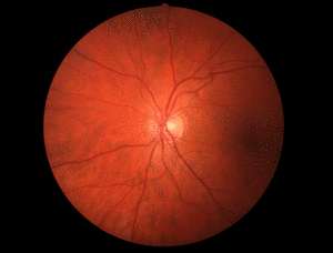
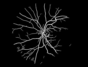
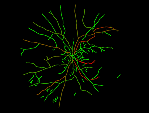
The following images show an example of the tortuosity visualisation:
The original RGB image is segmented to gain a binary image, showing only the vessel tree. The vessels in the binary image are tracked and separated from eachother. The tortuosity of each vessel is calculated independently, and visualized in a color coded image, where the green color shows vessels with normal tortuosity, and the red one the vessels with high tortuosity.
 |  |  |
Attila Budai, Joachim Hornegger, Georg Michelson: Multiscale Blood Vessel Segmentation in Retinal Fundus Images In: Meinzer, Hans-Peter; Deserno, Thomas Martin; Handels, Heinz; Tolxdorff, Thomas (Eds.) Bildverarbeitung für die Medizin 2010 - Algorithmen, Systeme, Anwendungen, (Bildverarbeitung für die Medizin 2010 - Algorithmen, Systeme, Anwendungen, Aachen 14-16.3.2010) Heidelberg : Springer 2010, pp. 261-265 - ISBN 978-3-642-11967-5
Attila Budai, Joachim Hornegger, Georg Michelson: Vessel Segmentation on Retinal Fundus Images: at The Association of Research in Vision and Ophthalmology (Konferenz) in Fort Lauderdale, FL, USA (03.05.2009)