
Invasive cardiac angiography (catheterization) is still the standard in clinical practice for diagnosing coronary artery disease (CAD) but it involves a high amount of risk and cost. New generations of CT scanners can acquire high-quality Images of coronary arteries which allow for an accurate identification and delineation of stenoses. Recently, computational flow Dynamics simulation has been applied to coronary blood flow using geometric lumen models extracted from CT angiography (CTA). The computed pressure drop at stenoses proved to be indicative for ischemia-causing lesions, leading to non-invasive Fractional Flow Reserve derived from CTA. Since the diagnostic value of non-invasive procedures for diagnosing CAD relies on an accurate extraction of the lumen, a precise segmentation of the coronary arteries is crucial. As manual segmentation is tedious, time-consuming and subjective, automatic procedures are desirable.
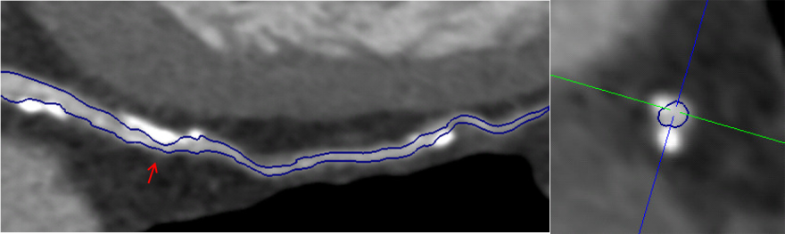
We present a novel fully-automatic method to accurately segment the lumen of coronary arteries in the presence of calcified and noncalcified plaque. Our segmentation framework is based on three main steps: boundary detection, calcium exclusion and surface optimization. A learning-based boundary detector enables a robust lumen contour detection via dense ray-casting. The exclusion of calcified plaque is assured through a novel calcium exclusion technique which allows us to accurately capture stenoses of diseased arteries. The boundary detection results are incorporated into a closed set formulation whose minimization yields an optimized lumen surface. On standardized tests with clinical data, a segmentation accuracy is achieved which is comparable to clinical experts and superior to current automatic methods.