Contact
 +49 9131 85 27894
 +49 9131 85 27270

Address
Chair of Computer Science 5 (Pattern Recognition)
Martensstraße 3
91058 Erlangen
Germany
Powered by


Sebastian Käppler M. Sc.
Alumnus of the Pattern Recognition Lab of the Friedrich-Alexander-Universität Erlangen-Nürnberg
Projects
S. Käppler, M. Seifert, F. Horn, G. Pelzer, J. Rieger, T. Michel, A. Maier, G. Anton, C. Riess
-
Grating-based Talbot-Lau interferometers are a popular choice for phase-contrast X-ray acquisitions. Here, an air reference scan has to be acquired prior to an object scan. This particularly complicates acquisition of large objects: large objects are tiled into multiple scans due to the small field of view of current gratings. However, phase reference drifts occurring between these scans may require to repeatedly move the object in and out of the X-ray beam to update the reference information.
We developed an image processing technique that completely removes the need for phase reference scans in tiled acquisitions. We estimate the reference from object scans using a tailored iterated robust regression, using a novel efficient optimizer. Our evaluation indicates that the estimated reference is not only close to the acquired reference but also improves the final image quality. We hypothesize that this is because we mitigate errors that are introduced when actually acquiring the reference phase. Phase-contrast imaging of larger objects may benefit from computational estimation of phase reference data due to reduced scanning complexity and improved image quality.
Journal ArticlesMedical Physics, vol. 44, no. 5, pp. 1886-1898, 2017 (BiBTeX, Who cited this?) -
Differential phase contrast image:
raw data, data processed with previous method
and proposed method.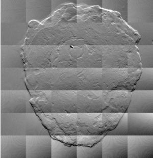
S. Käppler, F. Bayer, T. Weber, A. Maier, G. Anton, J. Hornegger, C. Riess et al.
-
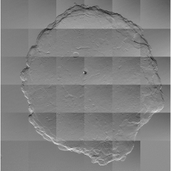
Grating-based X-ray dark-field imaging is a new imaging modality. It allows the visualization of structures at micrometer scale due to small-angle scattering of the X-ray beam. However, reading dark-field images is challenging as absorption and edge-diffraction effects also contribute to the dark-field signal, without adding diagnostic value. In this paper, we present a novel -- and to our knowledge the first -- algorithm for isolating small-angle scattering in dark-field images, which greatly improves their interpretability. To this end, our algorithm utilizes the information available from the absorption and differential phase images to identify clinically irrelevant contributions to the dark-field image. Experimental results on phantom and ex-vivo breast data promise a greatly enhanced diagnostic value of dark-field images.
Articles in Conference ProceedingsMedical Image Computing and Computer-Assisted Intervention -- MICCAI 2014 (International Conference on Medical Image Computing and Computer-Assisted Intervention), Boston, MA, USA, 15.09.2014, pp. 170-177, 2014, ISBN 978-3-319-10404-1 (BiBTeX, Who cited this?) -
Dark-field image of an ex-vivo human breast, before and after processing. 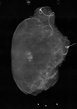
S. Käppler, J. Wandner, T. Weber, A. Maier, G. Anton, J. Hornegger, C. Riess
-
Grating-based differential phase-contrast X-ray is a novel imaging modality with excellent soft-tissue contrast. Besides standard X-ray attenuation, it provides complementary information on the differential phase shift and the dark-field signal, which reveals structure variations at (sub-)micron scale. Current experimental setups suffer from a narrow field of view of 2-4cm. Thus, multiple exposures have to be stitched together to image larger objects. However, individual exposures are inherently affected by intensity variations, such that tiling artifacts corrupt the stitched projection. These artifacts are most severe in the differential phase image and highly impact their diagnostic value.
To address this issue, we propose a novel optimization-based algorithm for fully compensating these tiling artifacts. Our algorithm estimates a smooth bias field for each individual exposure with a global objective function that minimizes the intensity distortion within and across different tiles in the projection. Compared to a currently widely used heuristic, our algorithm leverages the information available from all exposures to estimate the individual bias fields. The evaluation shows the superiority of the proposed algorithm, as it produces bias-free images. To our knowledge, this is the first bias correction algorithm for differential phase images that yields images with nearly imperceptible transitions between individual exposures.
Articles in Conference Proceedings2014 IEEE Nuclear Science Symposium and Medical Imaging Conference Record (NSS/MIC) ((IEEE Nuclear Science Symposium and Medical Imaging Conference (NSS/MIC) 2014)), Seattle, WA, USA, 08.11.2014, pp. 00-00, 2014 (BiBTeX, Who cited this?) -
Differential phase contrast image of an ex-vivo human breast, before and after processing.