
Contact
 +49 9131 85 27891
 +49 9131 85 27270

Address
Chair of Computer Science 5 (Pattern Recognition)
Martensstraße 3
91058 Erlangen
Germany
Powered by


Dr.-Ing. Yan Xia
Alumnus of the Pattern Recognition Lab of the Friedrich-Alexander-Universität Erlangen-Nürnberg
Region of interest reconstruction
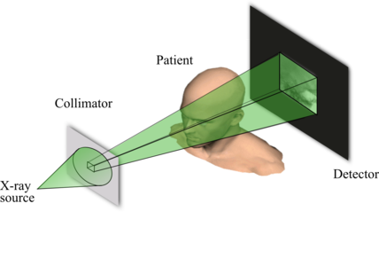
In some clinical applications and workflows (e.g. examination of deployed stents or coils after the intervention, cochlear implants, and needle biopsy) only a small portion of the whole patient may be of diagnostic interest, which enables the idea of utilizing X-ray beam collimation to laterally and axially block radiation during CT examination. Fig. 1 exemplarily shows the typical setup for ROI scan.
 |
ROI imaging
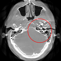
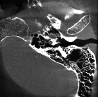
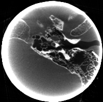
This ROI scan considerably reduces radiation dose to the patient due to the reduction in field of view (FOV), as illustrated in Figs. 2-4. Therefore, the corresponding ROI reconstruction emerges. It is well-suited for imaging guidance: 3D information is needed only in a small desired patient region without exposure of too much X-ray dose.
 |
 |
 |
Dynamic collimation
To track the defined 3D diagnostic region of interest, the collimator is controlled to move to an arbitrary position (normally asymmetric) in each projection. The dynamic scanning allows the acquisition of convex shapes around any point in the volume but the adjustment of the collimator is needed in each projection view for tracking the defined 3D ROI.
Reconstruction results
 |
 |
 |