
Contact


Address
Chair of Computer Science 5 (Pattern Recognition)
Germany
Powered by

Dr.-Ing. Andreas Fieselmann
Alumnus of the Pattern Recognition Lab of the Friedrich-Alexander-Universität Erlangen-Nürnberg
Interventional 4-D Tissue Perfusion Imaging with C-arm CT
1. Overview
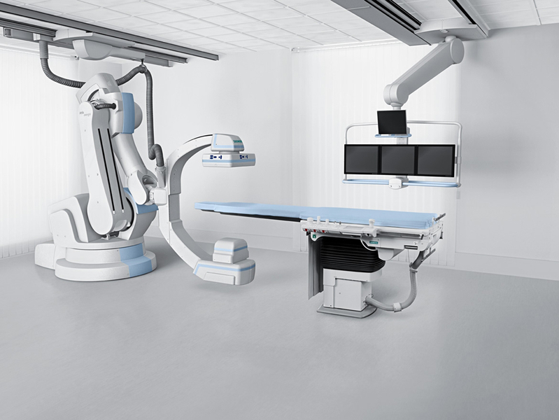
In this project, we investigate the use of a C-arm angiography system capable of CT-like imaging (C-arm CT, Fig. 1) to measure tissue perfusion. Tissue perfusion information, such as cerebral blood flow (CBF), can be used for stroke diagnosis and to guide further stroke treatment. While CT and MR-based perfusion imaging is well established perfusion imaging in the interventional suite using a C-arm CT is not available yet.
C-arm-CT-based perfusion imaging directly before the intervention - the state of perfusion may have changed after the initial CT/MR perfusion exam - or during the intervention - to determine the treatment endpoint - could optimize current stroke therapy procedures.
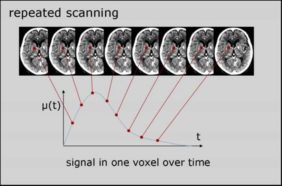
During perfusion imaging a region of interest is imaged several times at short intervals to study the flow of a previously injected bolus of contrast agent (Fig. 2). Using a C-arm CT for perfusion imaging faces certain challenges. In contrast to conventional CT scanners, the acquisition time for one data set is much longer (3-5 seconds compared to less than 1 second in CT) and thus the sample rate for the contrast flow is comparably low. Furthermore, inconsistencies in the projection images - due to the changing attenuation values from the contrast agent during the acquisition time - may cause reconstruction artifacts.
This project is in close collaboration with Siemens AG, Healthcare Sector, Angiography & Interventional X-Ray Systems, and the Department of Radiology, Stanford University, USA.
 |
 |
2. Research Projects
2.1 C-arm CT Perfusion Imaging Using Interleaved Scanning and Partial Reconstruction Interpolation
In this research project, we investigate specialized scan protocols to increase temporal sampling of the dynamic perfusion time-attenuation curves with current C-arm CT systems. These scan protocols employ several multi-rotational sequences where one multi-rotational sequence consists of several forward and backward rotations of the C-arm. There is a new contrast bolus injection, at a different relative delay time with respect to the start of the scan, for each sequence. Furthermore, we apply adapted reconstruction algorithms to optimally use the data from the interleaved scan protocols. See publications [1, 4, 8, 9, 10] for details.
2.2 Analysis and Reduction of FBP Artifacts Due to Time-Varying Attenuation Values
Filtered-backprojection-(FBP)-based reconstruction algorithms assume constant attenuation values during the acquisition of the projection data. If this assumption is violated reconstruction artifacts can arise. For example, in perfusion imaging with C-arm CT systems - which have acquisition times of several seconds - the (intentional) contrast flow can cause this violation. In this research project, we derive a formal description of these artifacts which can then be used to assess the magnitude of these artifacts and to reduce these artifacts by optimizing reconstruction parameters or developing new reconstruction approaches. See publication [2, 6] for details.
2.3 Contrast Bolus Measurement From 2-D Projection Data
Perfusion imaging requires a contrast bolus injection which, in CT and MR perfusion exams, is administered intra-venously. In C-arm CT perfusion imaging an intra-arterial contrast bolus injection at the aortic arch could be possible, to increase the SNR of the measured time-attenuation curves, because the patient is actually located in the interventional suite. Uniform contras bolus flow into both carotids is necessary for comparison of perfusion between the left and right hemispheres. In this research project, we investigate automatic methods to measure the distribution of the contrast flow into the carotid arteries to assess and optimize the injection catheter location. See publication [5] for details.
2.4 Evaluation of C-arm CT Image Quality With Respect to Perfusion Imaging
The focus of this research project is to evaluate the image quality of current C-arm CT systems with respect to dynamic perfusion imaging. In particular, we investigate the relationship between iodine concentration and measured Hounsfield values. See publication [7] for details.
3. Publications
Journal Articles
| [1] | A. Fieselmann, A. Ganguly, Y. Deuerling-Zheng, M. Zellerhoff, C. Rohkohl, J. Boese, J. Hornegger, R. Fahrig. Interventional 4-D C-Arm CT Perfusion Imaging Using Interleaved Scanning and Partial Reconstruction Interpolation. IEEE Transactions on Medical Imaging, 31(4), pages 892-906, 2012. (pdf, link) |
| [2] | A. Fieselmann, F. Dennerlein, Y. Deuerling-Zheng, J. Boese, R. Fahrig, and J. Hornegger. A model for filtered backprojection reconstruction artifacts due to time-varying attenuation values in perfusion C-arm CT. Physics in Medicine and Biology, 56(12), pages 3701-3717, 2011. (pdf, link) |
| [3] | A. Fieselmann, M. Kowarschik, A. Ganguly, J. Hornegger, and R. Fahrig. Deconvolution-based CT and MR brain perfusion measurement: Theoretical model revisited and practical implementation details. International Journal of Biomedical Imaging, vol. 2011, article ID 467563, 20 pages, 2011. (pdf, link) |
| [4] | A. Ganguly, A. Fieselmann, M. Marks, J. Rosenberg, J. Boese, Y. Deuerling-Zheng, M. Straka, G. Zaharchuk, R. Bammer, and R. Fahrig. Cerebral CT perfusion using an interventional C-arm imaging system: cerebral blood flow measurements. American Journal of Neuroradiology, 32(8), pages 1525-1531, 2011. (pdf, link) |
Conference Proceedings
| [5] | A. Fieselmann, A. Ganguly, Y. Deuerling-Zheng, J. Boese, J. Hornegger, and R. Fahrig. Automatic measurement of contrast bolus distribution in carotid arteries using a C-arm angiography system to support interventional perfusion imaging. In: Proc. SPIE Medical Imaging 2011: Visualization, Image-Guided Procedures, and Modeling, pages 79641W1-6, Orlando, USA, 2011. (pdf, link) |
| [6] | A. Fieselmann, F. Dennerlein, Y. Deuerling-Zheng, J. Boese, R. Fahrig, and J. Hornegger. A filter model to analyze reconstruction artifacts in perfusion C-arm CT. In: Proc. IEEE 2010 Nuclear Science Symposium and Medical Imaging Conference, pages 2239-2242, Knoxville, USA. 2010. (pdf, link) |
| [7] | A. Fieselmann, A. Ganguly, Y. Deuerling-Zheng, J. Boese, R. Fahrig, and J. Hornegger. Using a C-arm CT for interventional perfusion imaging: a phantom study to measure linearity between iodine concentration and Hounsfield values. In: Medizinische Physik 2010, Freiburg i. Br., Germany, 2010. (pdf, link) |
| [8] | A. Fieselmann, A. Ganguly, Y. Deuerling-Zheng, M. Zellerhoff, J. Boese, J. Hornegger, and R. Fahrig. A dynamic reconstruction approach for cerebral blood flow quantification with an interventional C-arm CT. In: Proc. IEEE International Symposium on Biomedical Imaging 2010, pages 53-56, Rotterdam, The Netherlands, 2010. (pdf, link) |
| [9] | A. Ganguly, A. Fieselmann, J. Boese, C. Rohkohl, J. Hornegger, and R. Fahrig. Evaluating the feasibility of C-arm CT for brain perfusion imaging: an in vitro study, In: Proc. SPIE Medical Imaging 2010: Visualization, Image-Guided Procedures, and Modeling, pages 76250K1-8, San Diego, USA, 2010. (pdf, link) |
| [10] | A. Fieselmann, A. Ganguly, Y. Deuerling-Zheng, M. Zellerhoff, M. Marks, J. Boese, and R. Fahrig. Volume cerebral blood flow (CBF) measurement using an interventional ceiling-mounted C-arm angiography system, In: Insights Into Imaging 2010 (European Congress of Radiology 2010 Book of Abstracts), page S186, Vienna, Austria, 2010. (pdf, link) |
Patents
| [11] | J. Boese, Y. Deuerling-Zheng, A. Fieselmann, and M. Zellerhoff. Verfahren zur Bestimmung der arteriellen Inputfunktion für Perfusionsmessungen und C-Bogen Röntgengerät (Measurement of arterial input function, during perfusion, uses two-dimensional X-ray projections and mask X-ray projections to compute a volume image) , German Patent DE102009004184B3. (pdf, link) |