
Contact


Address
Chair of Computer Science 5 (Pattern Recognition)
Martensstrasse 3
91058 Erlangen
Germany
Powered by

Dr.-Ing. Alexander Brost
Alumnus of the Pattern Recognition Lab of the Friedrich-Alexander-Universität Erlangen-Nürnberg
Alexander wins Klee-Preis 2013
Every year, the German Society for Biomedical Engineering (DGBMT) in cooperation with the Stiftung Familie Klee awards the DGBMT Prize of the Stiftung Familie Klee to support young talented researchers. This year, the award goes to Dr.-Ing. Alexander Brost for his dissertation “Image Processing for Fluoroscopy Guided Atrial Fibrillation Ablation Procedures”. Alexander wrote his thesis at the Pattern Recognition Lab of Prof. Dr.-Ing. Joachim Hornegger at the Friedrich-Alexander-University Erlangen-Nuremberg. After his graduation, he accepted a position at Stanford University, where he now works on image processing for magnetic resonance imaging for stroke and tumor patients. The official ceremony will be held during the annual meeting of the DGBMT in Graz, Austria, September 19 to 21.
(More information here: http://www.vde.com/de/fg/dgbmt/ehrungen-preise/seiten/klee-preisneu.aspx)

Alexander was a Postdoctoral Research Fellow
at the Center for Quantitative Neuroimaging,
Stanford University, Stanford, CA, USA, from June 2012 to June 2013.
 http://neuroquant.stanford.edu
- Image Processing
- Diffusion MRI
- Perfusion MRI
- Image Registration
- Image Guidance for Interventional Procedures
- Motion Compensation
- Catheter Tracking
- Object Reconstruction from Two Views
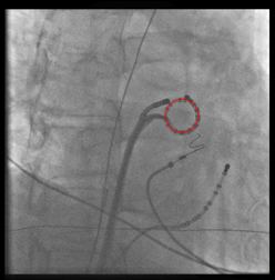
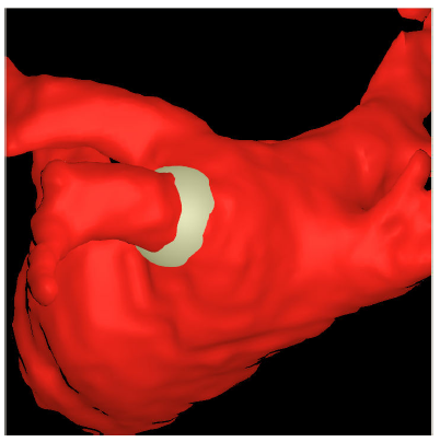
Motion compensation is facilitated by tracking a circumferential mapping catheter. This catheter is stably fixed at the ostium of the pulmonary vein considered for ablation. Our goal is to provide physicians a 3-D overlay rendered from diagnostic images such as CT or MRI to guide their procedures.
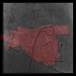
In fluoroscopic guided electrophysiology procedures, only 2-D projection images are available. To provide the physician 3-D information about the catheter positions, we reconstruct catheters from two views.
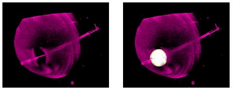 |
Atrial Fibrillation Ablation Planning Tool (AFiT)
Cryo-balloon catheter are so-called single-shot-devices. Our Atrial Fibrillation Ablation Planning Tool (AFiT) provides physicians a first planning tool for such procedures.
Room: 09.132
Telephone: +49 9131 85 27799
e-mail: Alexander.Brost(at)informatik.uni-erlangen.de